Biodiversity Heritage Library - Program news and collection highlights from BHL
  • Home
  • News
  • Featured Books
    • All Featured Books
    • Book of the Month Series
    • BHL at 20
  • User Stories
  • Campaigns
    • Fossil Stories
    • Garden Stories
    • Monsters Are Real
    • Page Frights
    • Her Natural History
    • Earth Optimism 2020
  • Tech Blog
  • Visit BHL
Home
News
Featured Books
    All Featured Books
    Book of the Month Series
    BHL at 20
User Stories
Campaigns
    Fossil Stories
    Garden Stories
    Monsters Are Real
    Page Frights
    Her Natural History
    Earth Optimism 2020
Tech Blog
Visit BHL
  • Home
  • News
  • Featured Books
    • All Featured Books
    • Book of the Month Series
    • BHL at 20
  • User Stories
  • Campaigns
    • Fossil Stories
    • Garden Stories
    • Monsters Are Real
    • Page Frights
    • Her Natural History
    • Earth Optimism 2020
  • Tech Blog
  • Visit BHL
Biodiversity Heritage Library - Program news and collection highlights from BHL
Blog Reel, User Stories

Finding Refuge in the Library: How BHL Inspired the Mycological Book Club

Screenshot of the Twitter profile of @MycoBookClub.

On 23 March 2020, the U.K. went into its first national lockdown in response to the COVID-19 pandemic. Social distancing and teleworking became the norm, and the walls of our homes became, in many ways, the boundaries of our individual worlds.

Amateur mycologist Clare Blencowe was eager to find a positive distraction from the realities of life during a pandemic. The Biodiversity Heritage Library’s open access collections, which for Blencowe had become a welcome refuge from the continuous onslaught of negative news articles, now became the inspiration for a new, socially distanced way to connect with other fungi lovers—in the form of “The Mycological Book Club”, an online, Twitter-based book club with a particular focus on open access literature.
#MycoBookClub logo fungi growing out of a book

#MycoBookClub logo, designed by illustrator Kelsey Sconberg of Mossy Stone Media.

“We were at the beginning of a national lockdown,” recalls Blencowe. “All social events were cancelled, and I found myself endlessly scrolling through increasingly doom-laden news, feeling increasingly doom-y. #MycoBookClub started as a simple distraction, an opportunity for a shared experience: people reading the same book and discussing it on Twitter at an allotted time.”
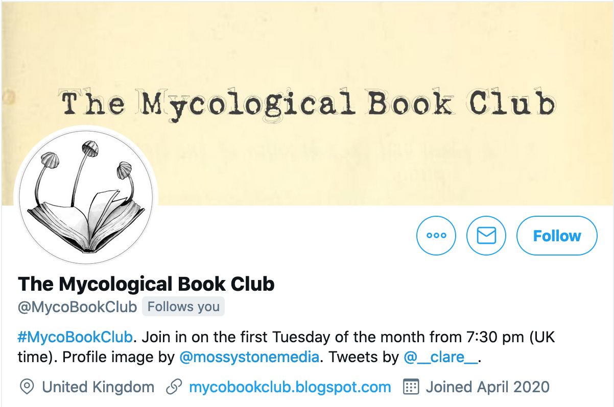
Screenshot of the Twitter profile of @MycoBookClub.

@MycoBookClub on Twitter.

Blencowe brought her idea to life by founding the @MycoBookClub Twitter account, establishing the #MycoBookClub hashtag to facilitate discussion, and setting a recurring date and time for book club meetings—the first Tuesday of the month at 7:30pm (GMT) on Twitter. She then scheduled the first #MycoBookClub meeting for 5 May 2020.

That first meeting, which brought together just a handful of followers, was dedicated to Mordecai Cubitt Cooke’s Rust, Smut, Mildew & Mould: An Introduction to the Study of Microscopic Fungi (Fifth ed. 1886), which club members accessed through BHL.

Welcome to #MycoBookClub, folks! It’s a pleasant evening here at @MycoBookClub headquarters in Sussex, England.

We’re talking about ‘MICROSCOPIC FUNGI: Rust, Smut, Mildew & Mould’ by M.C. Cooke (1886). You can find it at: https://t.co/7WO005ZS5Q pic.twitter.com/GxWzmYhnxg

— The Mycological Book Club (@MycoBookClub) May 5, 2020

With passages celebrating the beauty and wonder of the often unheeded nature in our own backyards and neighborhoods, the title was a fitting—and motivating—choice for an audience confined to their homes and local natural surroundings.

As Cooke writes,

“We might traverse the primeval forests of the new world, and explore the unknown regions of the old, and not encounter so much to excite our admiration, or cause our wonder, as lies about our feet at home; marvels which we tread beneath our feet, or kick from our path, because they appear to be only rotten sticks, withered grass, and decaying leaves.”

“Cooke’s writing provided inspiration for enjoying nature near home,” shares Blencowe. “I felt myself lifted by his soaring rhetoric.”
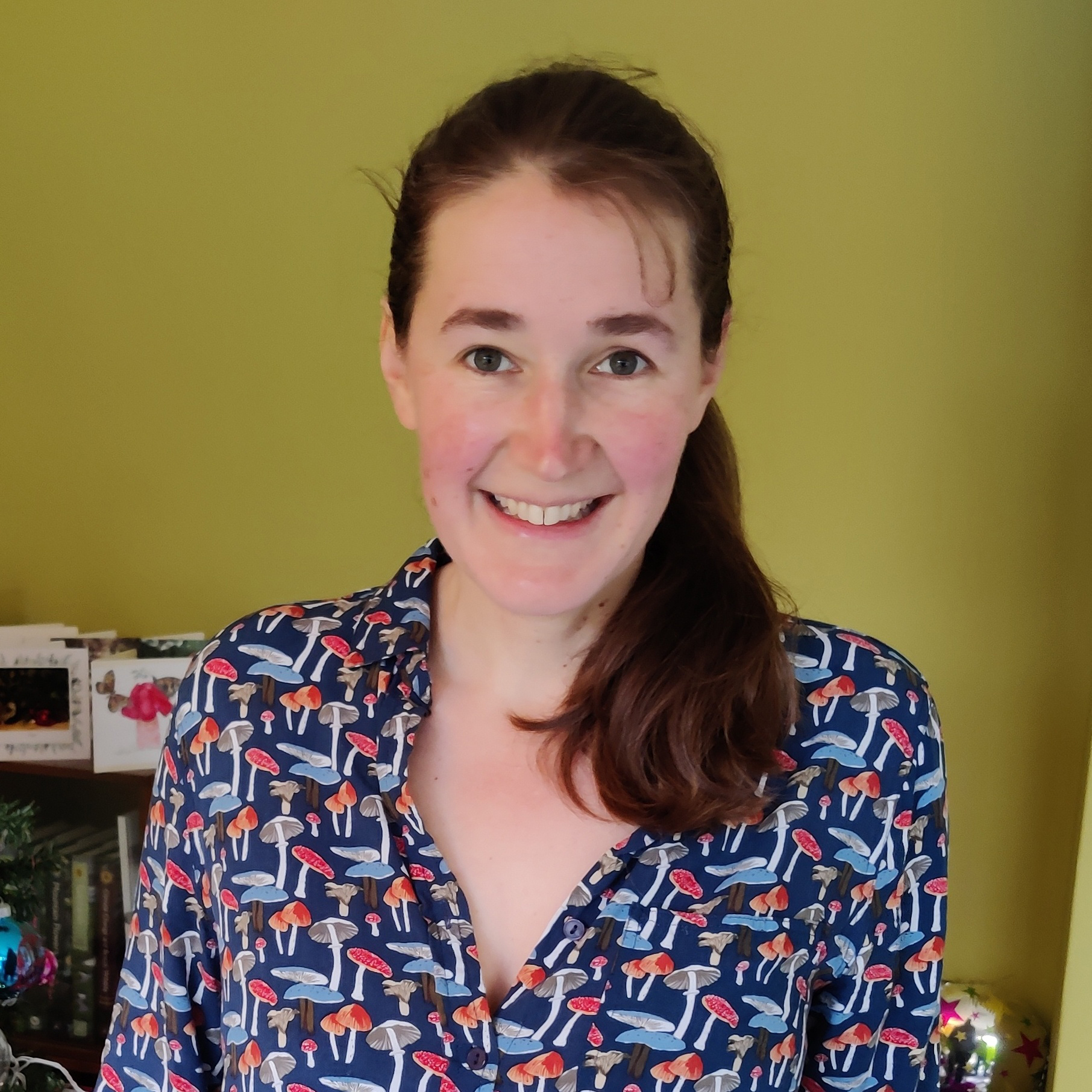
Photo of a human with long brown hair in a shirt with fungi images on it.

Amateur mycologist Clare Blencowe, founder of the #MycoBookClub. Photo courtesy of Clare Blencowe.

#MycoBookClub has allowed Blencowe to share and grow her interest in mycology—an interest that began in earnest about five years ago when a new job as manager of the Sussex Biodiversity Record Centre, and the much shorter commute that came with it, provided her with newfound time to pursue her interest in fungi. While she considers mycology a hobby, her involvement in the field has grown over the years through membership in the British Mycological Society, where she was recently elected to their Field Mycology and Conservation Committee.

BHL helped nurture this love of mycology well before the birth of #MycoBookClub. Blencowe’s first in-depth interaction with the Library occurred in April 2019, when her discovery of a small brown fungus cup in the woods led her to seek confirmation of its identity—which she believed to be the Anemone Cup Dumontinia tuberosa—by posting to the British Mycological Society Facebook group. UK-based mycologist Lukas Large responded to her post with the link to a color plate for comparison from Émile Boudier’s Icones Mycologicæ (1905-1910) in BHL.
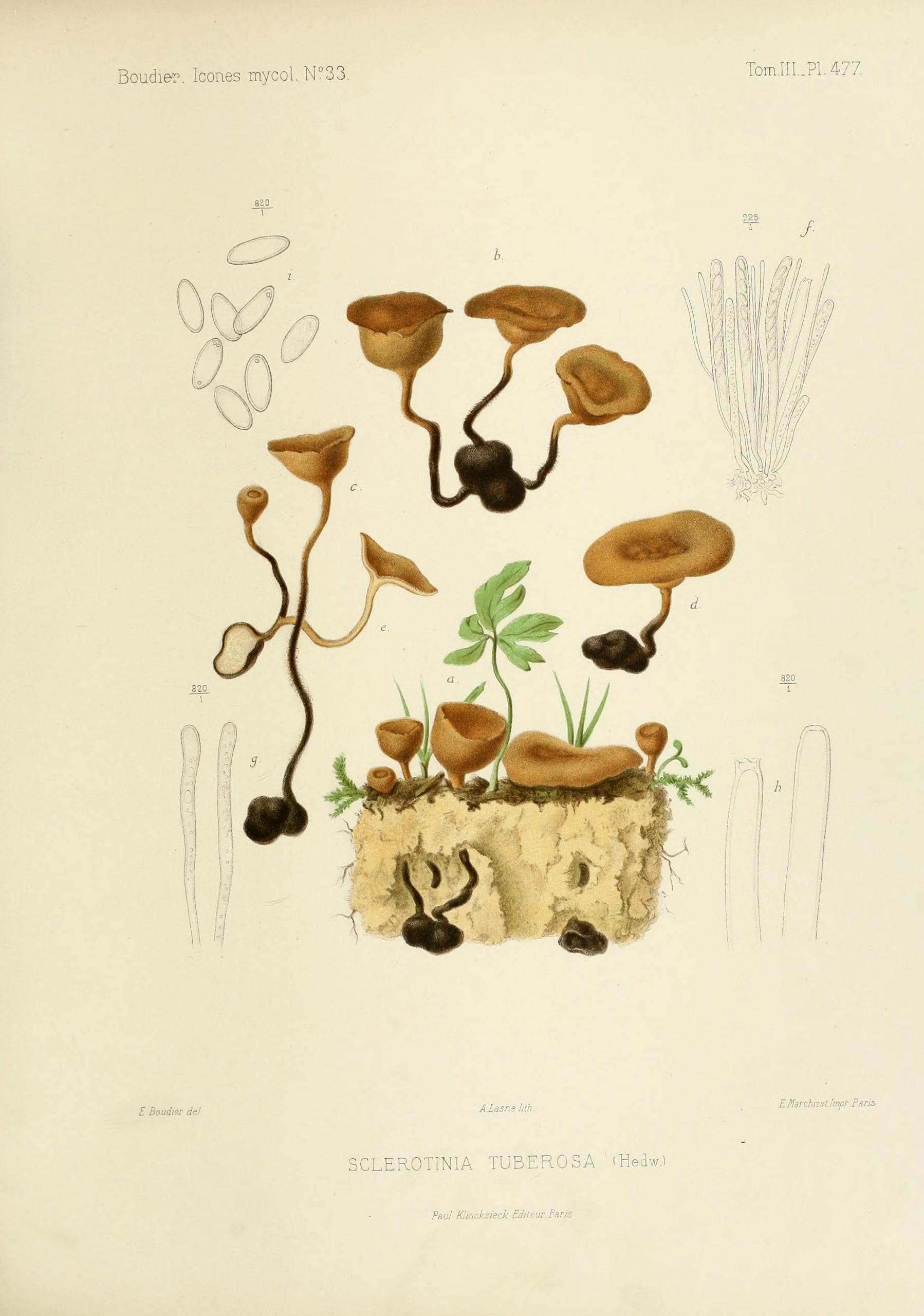
Illustration of fungi.

Illustration of Dumontinia tuberosa, plate 477. Boudier, Émile. Icones Mycologicæ (1905-1910), v.3. Art by Boudier. Contributed in BHL from the University Library, University of Illinois Urbana Champaign.

“The information contained in this beautiful colour plate far surpassed anything I had access to in my field guides at home,” shares Blencowe. “It helped me to understand the structure and features of this fungus I had in front of me, and spurred me on to take a closer look at it under the microscope. That experience really opened my eyes to what a useful resource the books in BHL can be: to inform observation and help identify species in a present day context. Sure, species concepts and nomenclature may have moved on, but the detail with which mycologists such as Boudier recorded their observations is a lesson in itself.”

Since that eye-opening experience, Blencowe continues to turn to BHL as a resource to support her interests in mycology, reading titles online and downloading select pages and images as needed. Some of her favorite aspects of the Library include a good search functionality, user-friendly online reading and content downloading, high quality scans, clear copyright information, easily-shareable direct page links, and the image collections in Flickr. Since founding #MycoBookClub, she uses BHL almost every week.

We’ve talked already here at #MycoBookClub about M.C. Cooke, but tonight we’re also feasting our eyes on ‘Selecta Fungorum Carpologia’ by The Brothers Tulasne.

You can find their illustrations in the Biodiversity Heritage Library: https://t.co/DN4Hi816XE pic.twitter.com/buaU9fMTa4

— The Mycological Book Club (@MycoBookClub) June 2, 2020

“I love the little bookish details that are preserved in BHL: the gilt decoration on a publication’s cover, ornate book plates, library stamps, barely-decipherable pencil marks and blank pages uniquely speckled with foxing,” shares Blencowe. “The scanned pages hold so much more than simply the words and the illustrations. They do your soul good.”

A reminder that it’s #MycoBookClub night tonight, from 7:30pm UK time.

We’re trying out a theme this evening: illustration ?https://t.co/Bvx0nDHY08

— The Mycological Book Club (@MycoBookClub) November 3, 2020

In the months following the club’s first meeting in May 2020, #MycoBookClub continued to grow, amassing more followers and receiving attention in outlets like The British Mycological Society Newsletter and IMA Fungus. Blencowe established a “virtual bookshelf” to maintain a list of all the books that #MycoBookClub had discussed—many of which were accessed through BHL.
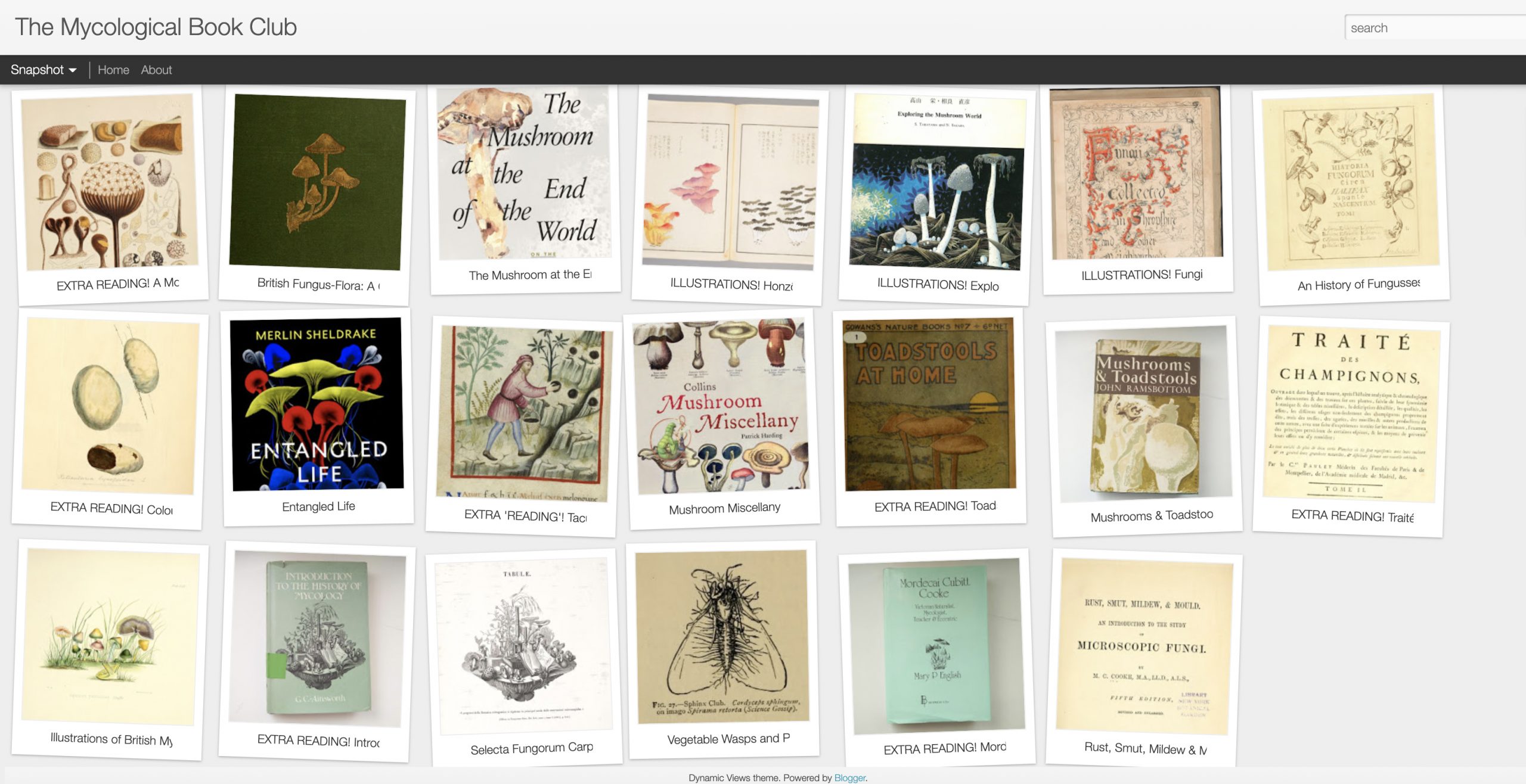
Screenshot of a virtual bookshelf with thumbnails of book covers.

#MycoBookClub “virtual bookshelf.” Source: https://mycobookclub.blogspot.com

“The more time I spend in the Library, the more amazed I am by the vast amount of literature it contains—and the neat way it is organised and presented,” lauds Blencowe. “BHL also provides the opportunity to take a deep dive into the source material for a fascinating glimpse into the machinery of global science in the 19th century.”

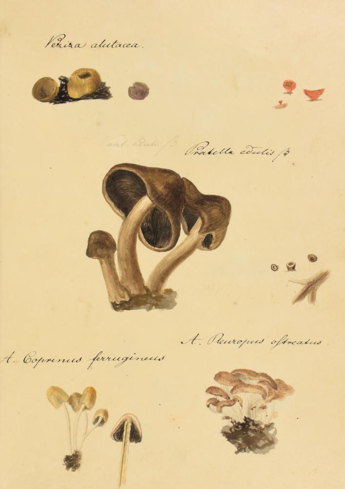
While she’s enjoyed many titles in BHL, a recent favorite is Anna Maria Hussey’s Illustrations of British Mycology (1847-55), which was the subject of the July 2020 #MycoBookClub meeting. Published under the name Mrs. T.J. Hussey, the two-volume work illustrates and describes species collected by Hussey and her sister, Frances Reed.

#MycoBookClub returns Tues 7 July, 7:30pm (UK time).

Thought we could take a broader look at the fungal kingdom this month with ‘Illustrations of British Mycology’ by Anna Maria Hussey (1847).

You can read it in the @BioDivLibrary, here: https://t.co/4BPXfDyWhd pic.twitter.com/icBcIRd1xM

— The Mycological Book Club (@MycoBookClub) June 7, 2020

“Hussey writes from the perspective of an enthusiastic amateur which I found both highly relatable and genuinely inspiring,” affirms Blencowe. “She writes in a chatty, conversational style which is peppered with fascinating contemporary details (such as her observation that Boletus subtomentosus has ‘the texture of a Limerick glove’) and timeless wisdom (‘life passes in gaining experience for ourselves…till at last we have acquired the wisdom to guide us safely…’).”
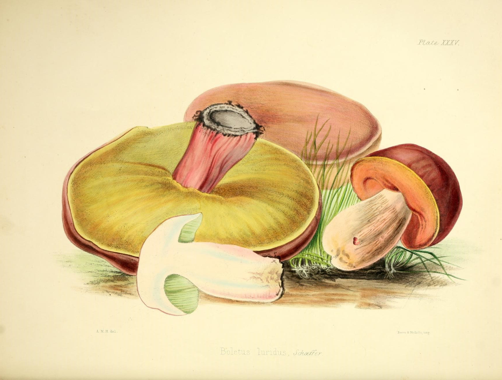
Illustration of fungi.

Illustration of Suillellus luridus, plate 35. Hussey, Anna Maria. Illustrations of British Mycology, ser.2 (1855). Art by Hussey. Contributed in BHL from The New York Botanical Garden, LuEsther T. Mertz Library. Explore all illustrations from Illustrations of British Mycology in the BHL Flickr.

Blencowe has gathered and shared via @MycoBookClub some of her favorite elements from Hussey’s work, including her approach to mycology, her discussion of fungus names, her commentary on fungi in nature, her descriptions of fungal habitats, and miscellaneous myco-chatter. Illustrations of British Mycologyis also noteworthy as a rare female-authored work in the bibliography of historic mycological literature.

“Browsing the historic biodiversity literature, it is very noticeable that certain perspectives predominate,” notes Blencowe. “The books are mostly written by men—white men. So it was a particular joy to come across Hussey’s book. I’m keen to explore ways that @MycoBookClub can provide a platform for different perspectives and respectfully champion Indigenous knowledge of fungi.”

While ours may still be a socially-distanced world, initiatives like #MycoBookClub remind us that there are still opportunities to connect with others, share our passions, and nurture a community. And while the news may still be filled with dire headlines, naturalists like Mordecai Cubitt Cooke and Anna Maria Hussey remind us that we can still find solace in nature close to home and in unexpected places, if we are just willing to look.

To quote Hussey: “The world is full of beauty that we pass by unheeded.”
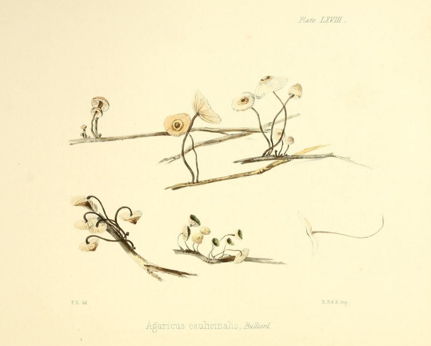
Illustration of fungi.

Illustration of Crinipellis scabella, plate 68. Hussey, Anna Maria. Illustrations of British Mycology, ser.1 (1847). Art by Frances Reed. Contributed in BHL from The New York Botanical Garden, LuEsther T. Mertz Library. This plate accompanies Hussey’s quote: “The world is full of beauty that we pass by unheeded.”

Everyone is welcome to join the #MycoBookClub. Follow @MycoBookClub for updates and meeting information, and join the discussion the first Tuesday of each month at 7:30pm (GMT) using the #MycoBookClub hashtag on Twitter.
February 1, 2021by michelle.underhill
Blog Reel, Campaigns, Her Natural History

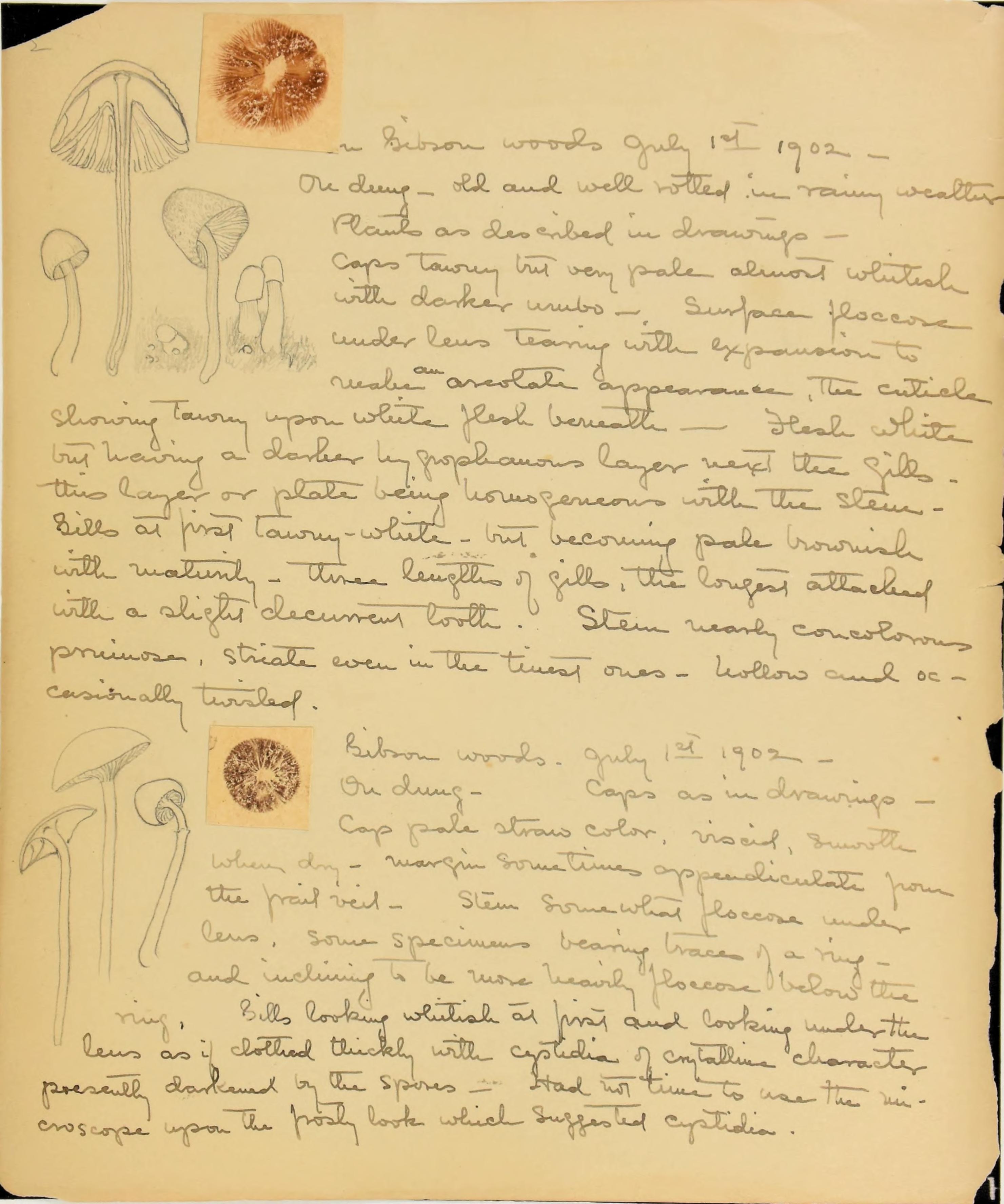
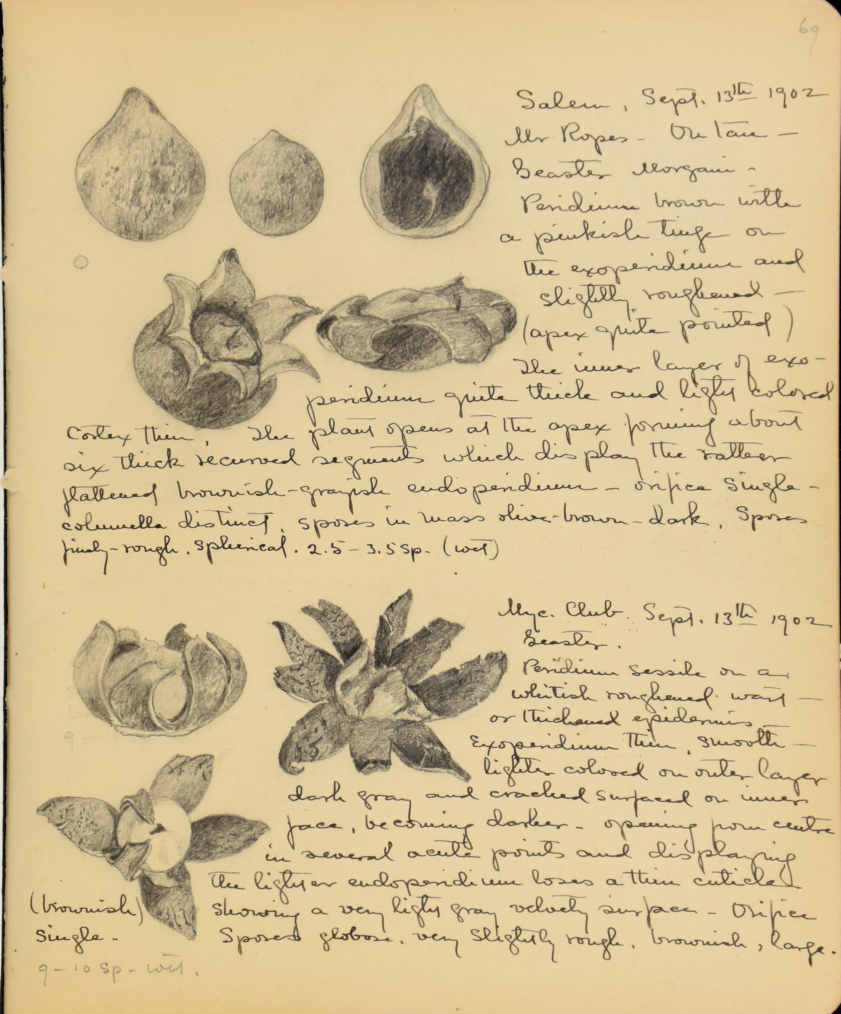
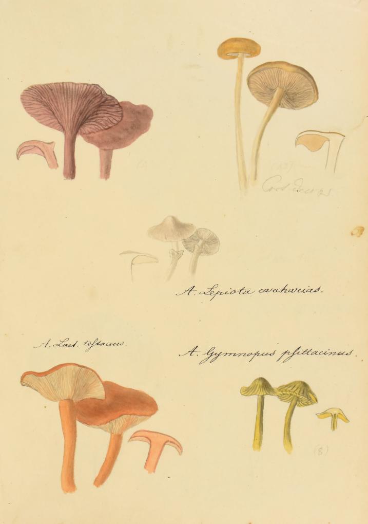
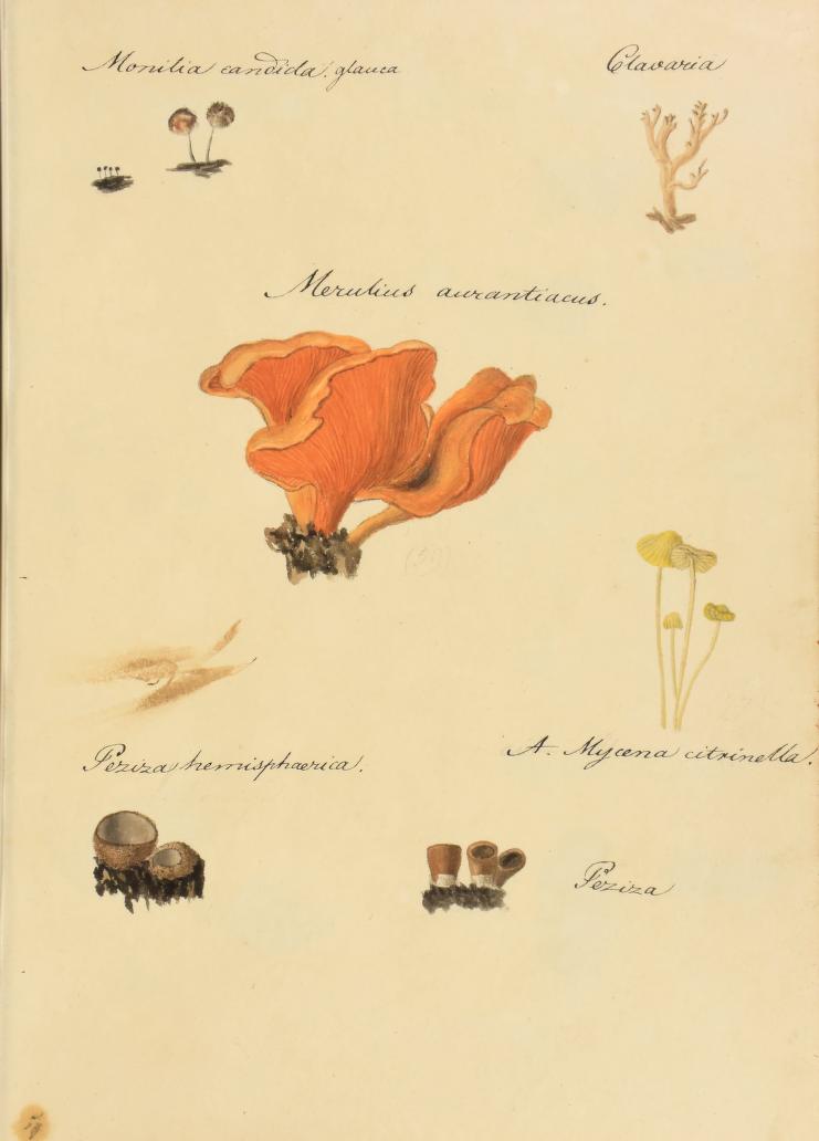
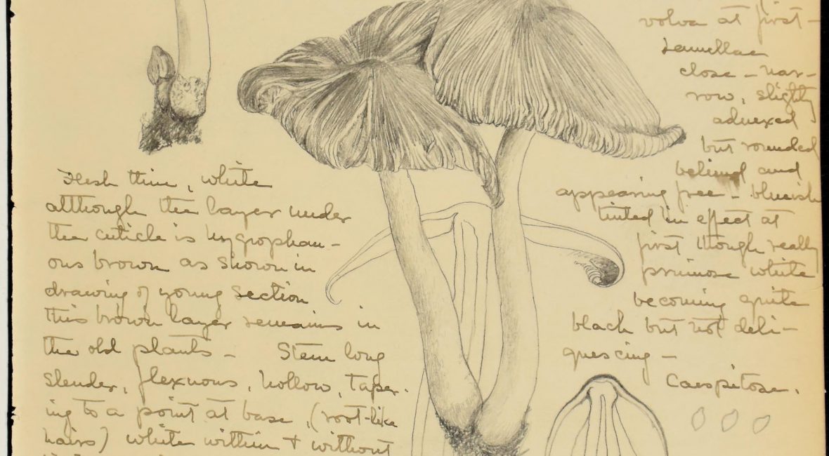
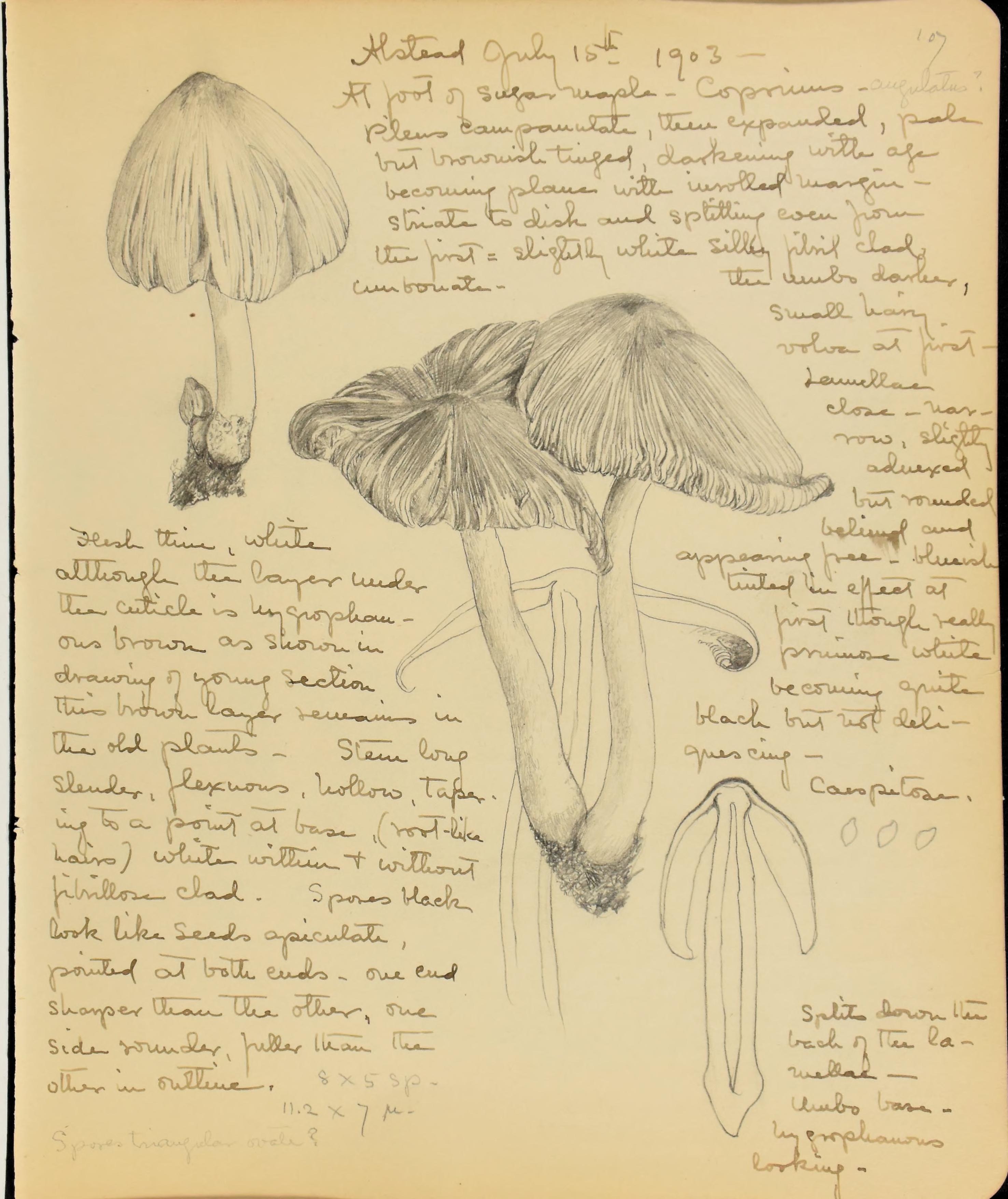
Henrietta Page sketchbook of fungi drawings

Page, Henrietta. [Sketchbook of drawings of fungi], volume II. Contributed in BHL Harvard University Herbarium, Botany Libraries.

The Harvard Botany Libraries recently acquired and digitized Henrietta Page’s [Sketchbook of drawings of fungi], volume II. The sketchbook was a gift from the Boston Mycological Club (BMC) where Page was a member and served on the executive committee for a time. She was also a member of the Boston Society of Natural History. The sketchbook mostly describes fungi collected during July and August when Page attended three-week long natural history classes held in Alstead, New Hampshire under the Alstead School of Natural History from 1899-1903. The mycology section was taught by Hollis Webster, secretary and co-founder of the BMC and Harvard graduate (class of 1884); phanerogamic botany by Merritt Lyndon Fernald; ornithology by Ralph Hoffmann; and physiography and biology by William Lusk Webster Field. The school ran from 1899-1904.

The sketchbook was originally unattributed, but Jason Karakehian (BMC archivist) discovered that the sketchbook connects to specimens that are now housed in the fungarium at New York Botanical Garden (NYBG). The specimens were originally part of the Alstead School of Natural history fungarium, which was integrated into the BMC fungarium after the Alstead School’s closing. Jason was able to attribute the sketchbook to Ms. Page by searching the NYBG’s online database of specimens for fungal collections made in Alstead in the years 1900-1903, which yielded a few collections whose labels referred to Henrietta Page by name and referenced specific pages in her sketchbook “Vol. II.”

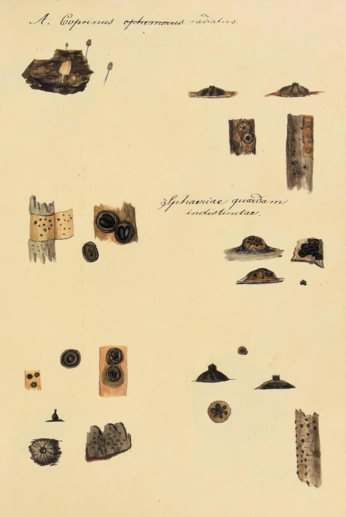
Page, Henrietta. [Sketchbook of drawings of fungi], volume II. Contributed in BHL Harvard University Herbarium, Botany Libraries.

There are a number of glass plate negatives, prints, and lantern slides of fungi in the BMC archive which were made by Hollis Webster in the 1902 and 1903 sessions of the School. These have been cleaned, organized, placed in archival negative envelopes, and listed in a spreadsheet. The specimens photographed do not appear to match those drawn by Ms. Page, but there is some overlap in taxa.

The sketchbook is a popular new acquisition and has already been used for several classes interested in field notes, women in science, and scientific illustrations.

Page, Henrietta. [Sketchbook of drawings of fungi], volume II. Contributed in BHL Harvard University Herbarium, Botany Libraries.

March 27, 2019by Joel Richard
Blog Reel, User Stories

Uncovering Mycological History…One Sketch at a Time

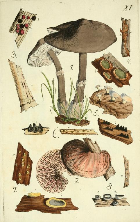
illustration of fungi

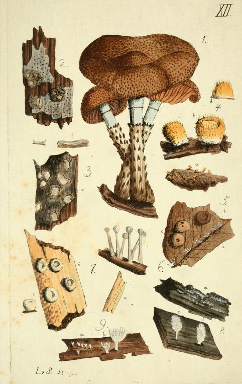
Plate 12. Schweinitz, Lewis David von. Icones fungorum Niskiensium. (ca.) 1798-1802. Contributed in BHL from Botany Libraries, Farlow Reference Library of Cryptogamic Botany, Harvard University.

In the archives of the Farlow Reference Library of Cryptogamic Botany at Harvard University, there is a curious volume of 249 original watercolors of fungi species bearing the title Icones fungorum Niskiensium.

Devoid of any creator identification, the provenance and historical significance of the volume seems shrouded in mystery. A cursory examination of the work reveals a collection of charming, but often incomplete, figures and a myriad of annotations in pencil and ink, some struck through, others revealing uncertainty over species identifications or recording observations on specimen quality or coloration.

illustration of fungi

Plate 47. Schweinitz, Lewis David von. Icones fungorum Niskiensium. (ca.) 1798-1802. Contributed in BHL from Botany Libraries, Farlow Reference Library of Cryptogamic Botany, Harvard University.

In short, there is little to suggest that this sketchbook is actually an important document in the history of mycological research.

The truth, however, is that this volume is the work of the “Father of North American mycology”, Lewis David von Schweinitz (1780-1834). It served him as a sketchbook that was used to inform his monumental Conspectus fungorum in Lusatiae Superioris agro Niskiensi crescentium, e methodo Persooniana (1805), co-authored with Johannes Baptista von Albertini (1769-1831) and documenting over 1,000 species of fungi from the Niesky, Germany region, including 100 published as new-to-science. The volume is also related to a collection of six other original watercolor volumes by Schweinitz dispersed throughout several American institutions.

Illustration of fungi

Plate 11. Conspectus fungorum in Lusatiae Superioris agro Niskiensi crescentium, e methodo Persooniana(1805). Figures based on drawings by Schweinitz. Icones fungorum Niskiensium served as a sketchbook to inform this work. Contributed to BHL by the LuEsther T. Mertz Library of The New York Botanical Garden.

The history of this volume, dubbed the Harvard Icones, and its relationship to the Conspectus and Schweinitz’s other watercolor volumes, was recently examined in-depth within the article “New Light on the Mycological Work of Lewis David von Schweinitz” (2018) by Jason M. Karakehian, William R. Burk and Donald H. Pfister, published in IMA Fungus: 9(1). The article presents research made possible in-part thanks to the digital copies of the Harvard Icones and Schweinitz’s other works available in the Biodiversity Heritage Library.

We recently explored this fascinating history in our Book of the Month series. The findings presented by Karakehian et al. underscore the importance of the Harvard Icones to the development of Schweinitz and Albertini’s research and the progression of mycology in the early 19th century.

“Our research is important because the volumes of watercolor illustrations produced by Schweinitz have been little known to mycologists who worked with fungi described by Albertini and Schweinitz,” explains Karakehian. “These high-quality illustrations can help to inform our concepts of some of these species, as often times early nineteenth century verbal descriptions can be short and somewhat vague.”

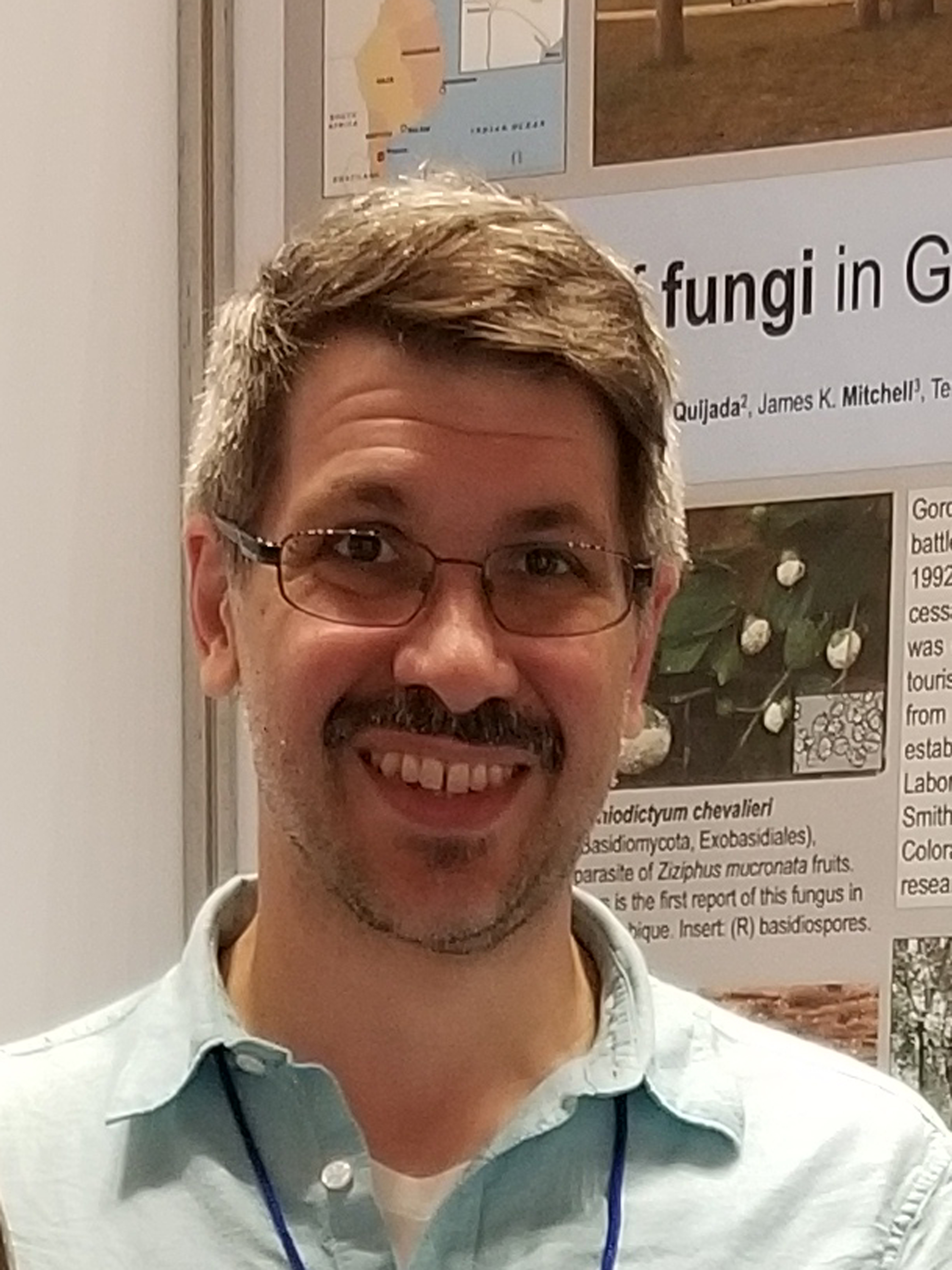
photo of a man in glasses and blue shirt

Jason M. Karakehian. Master’s degree candidate at the Harvard Extension School.

Jason M. Karakehian is a Master’s degree candidate at the Harvard Extension School, studying biology with a focus on mycology and the systematics of ascomycetes or “cup-fungi.” With a background in the visual arts, Karakehian’s interest in mycology stems from his career as an artist, when he began perusing illustrated books on fungi as a creative reference point for his large, outdoor sculptures exhibited in wooded environments. This inspired him to take a short class on fungi, join a local mycological club, and begin collecting and identifying mushrooms. Eventually, he gave up his art career and embraced mycology full-time.

Karakehian’s journey down the fungi path includes serving as the archivist for the Boston Mycological Club. The task of organizing the mass of artifacts and documents held by the archives necessitated research at other libraries and archives, such as the Reference Library of the Farlow Herbarium of Cryptogamic Botany at Harvard University. He was welcomed by the Herbarium and Library staff, including Professor Donald Pfister and Judy Warnement (Head Librarian of the Harvard Botany Libraries).

Karakehian’s work at the Boston Mycological Club and the relationships he developed as part of that process led to research on other topics in mycological history, especially Schweinitz’s fungi watercolors.

The Biodiversity Heritage Library played an essential role in that research.

“I don’t think that we could have done this work without regular access to BHL,” affirms Karakehian. “BHL is invaluable to us for the wide selection of classic mycological literature that we needed to reference in our research.”

illustration of fungi

Plate 23. Schweinitz, Lewis David von. Icones fungorum Niskiensium. (ca.) 1798-1802. Contributed in BHL from Botany Libraries, Farlow Reference Library of Cryptogamic Botany, Harvard University.

That research involved indexing the more than 1,000 species treated in the Conspectus and cross-referencing them to the species figured in Schweinitz’s watercolor volumes in order to examine the relationship between the works and understand how the archival materials informed Schweinitz’s research.

This work required regular access to Schweinitz’s Conspectus and other classic mycological publications, including Christiaan Hendrik Persoon’s Synopsis methodica fungorum (1801), which outlines a systematic framework for fungi that was employed by Schweinitz and Albertini.

“For both of these volumes, we had to go through them page by page to populate spreadsheets with data that we could then collate to make inferences about the relationship between them,” explains Karakehian. “This was greatly facilitated by having high-quality electronic versions of the volumes with OCR.”

Karakehian and his colleagues also created an index to the figures in the Harvard Icones, cross-referencing them to their treatment in the Conspectus, Persoon’s Synopsis, and Schweinitz’s six other watercolor volumes. This index was published as part of their IMA Fungus article.

illustration of fungi

Plate 41. Schweinitz, Lewis David von. Icones fungorum Niskiensium. (ca.) 1798-1802. Contributed in BHL from Botany Libraries, Farlow Reference Library of Cryptogamic Botany, Harvard University.

For Karakehian, BHL’s impact is about more than just providing online access to materials. It’s the quality of the available materials that really distinguishes BHL from other online repositories.

“BHL volumes are high-quality digitizations and I am never frustrated by missing pages, cut-off words, or poor-quality digitizations of delicate line drawings in figures,” lauds Karakehian. “I often think of BHL in contrast to Google’s digitization project that is easily searchable, but the digitizations are of such poor quality that one often needs to track down the original volume somehow to understand the print or the figure that you need.”

Karakehian and his colleagues were instrumental in advocating for Schweinitz’s archival materials to be made available online and for free to the public through BHL. This not only facilitates future research on these works, but also allows more people to explore and enjoy these archives and the history they represent.

“For us, BHL is the gold standard, and for our papers on Schweinitz’s watercolor illustrations, it was important to request that the archives holding these volumes digitize them and have them available in BHL to the public,” asserts Karakehian. “We were absolutely confident that the product would be usable and high-quality. It is a true gift that they are now freely available to users.”

illustration of fungi

Plate 13. Schweinitz, Lewis David von. Icones fungorum Niskiensium. (ca.) 1798-1802. Contributed in BHL from Botany Libraries, Farlow Reference Library of Cryptogamic Botany, Harvard University.

We are honored to host a digital copy of the Harvard Icones in BHL, and are grateful to the Botany Libraries, Farlow Reference Library of Cryptogamic Botany, Harvard University for digitizing the volume and making it freely available to the world. We’re also thrilled that Schweinitz’s other archival materials and the Conspectus are freely available in the collection, and extend our sincere thanks to the Library & Archives of the Academy of Natural Sciences of Drexel University, the Wilson Library of the University of North Carolina at Chapel Hill, and the LuEsther T. Mertz Library of The New York Botanical Garden for digitizing these works.

Thanks to these contributions from so many libraries, BHL was able to play a vital role in allowing Karakehian and his colleagues to provide new insight into the contributions of such an important figure in the history of mycology.

“Our work broadens our knowledge of the scientist and artist that Schweinitz was,” affirms Karakehian. “He was an accomplished and prolific mycological illustrator with a keen sense of depicting his subjects in ways that would convey the maximum of biologic information as well as capturing their natural beauty.”

That “natural beauty” is now freely available for everyone to enjoy through the Biodiversity Heritage Library.

November 8, 2018by michelle.underhill
Blog Reel, Featured Books

Unearthing Scientific History through Art: New Insights from the Archives of Lewis David von Schweinitz, the “Father of North American Mycology”

illustration of fungi

The information in this post is derived from the article “New Light on the Mycological Work of Lewis David von Schweinitz” (2018) by Jason M. Karakehian, William R. Burk and Donald H. Pfister, published in IMA Fungus: 9(1).

fungi illustration

Plate 12. Conspectus fungorum in Lusatiae Superioris agro Niskiensi crescentium, e methodo Persooniana (1805). Figures based on drawings by Schweinitz. Icones fungorum Niskiensium served as a sketchbook to inform this work. Contributed to BHL by the LuEsther T. Mertz Library of The New York Botanical Garden.

In 1805, the “Father of North American mycology”, Lewis David von Schweinitz (1780-1834), published an account of the fungi in Niesky, Germany with his friend and mentor, Johannes Baptista von Albertini (1769-1831). Documenting over 1,000 species, including 100 published as new-to-science, Conspectus fungorum in Lusatiae Superioris agro Niskiensi crescentium, e methodo Persooniana is still referenced to this day as a classic mycological text and ecological record.

The Conspectus is illustrated with 12 hand-colored plates based on drawings by Schweinitz, each featuring 6-10 figures of new species described. Whenever possible, Schweinitz based his drawings on fresh specimens, but when this was not an option, he referred to fungarium specimens or to a collection of earlier watercolors he’d created of representative specimens.

fungi illustrations

Plate 178. Fungorum Nieskiensium Icones. (ca.) 1802-1805. v. 3. Contributed in BHL from the Academy of Natural Sciences of Drexel University, Library & Archives.

Today, these watercolor volumes are dispersed between several American institutions and offer a wealth of insight into the history and development of the Conspectus. A five-volume set created (ca.) 1802-1805 and bearing the title Fungorum Nieskiensium Icones is distributed between the Archives of the Academy of Natural Sciences of Drexel University, Philadelphia (volumes 1-3 and 5) and the Rare Book Collection, Wilson Library, University of North Carolina, Chapel Hill (volume 4). Nearly every specimen featured in the Conspectus has a corresponding illustration in these 5-volumes.

A volume of original watercolors consisting of copies of figures from the 5-volume set, bearing the title Icones Fungorum and created (ca.) 1818-1826, is held by the Herbarium Library, University of Michigan, Ann Arbor, MI.

The earliest volume of original watercolors related to the Conspectus, however, is the Icones Fungorum Niskiensium, created (ca.) 1798-1802 and held in the archives of the Farlow Reference Library of Cryptogamic Botany at Harvard University. This volume (hereafter referred to as the Harvard Icones) served Schweinitz as a sketchbook of sorts, which he used to inform the final paintings in the 5-volume Fungorum Nieskiensium Icones and eventually the Conspectus itself.

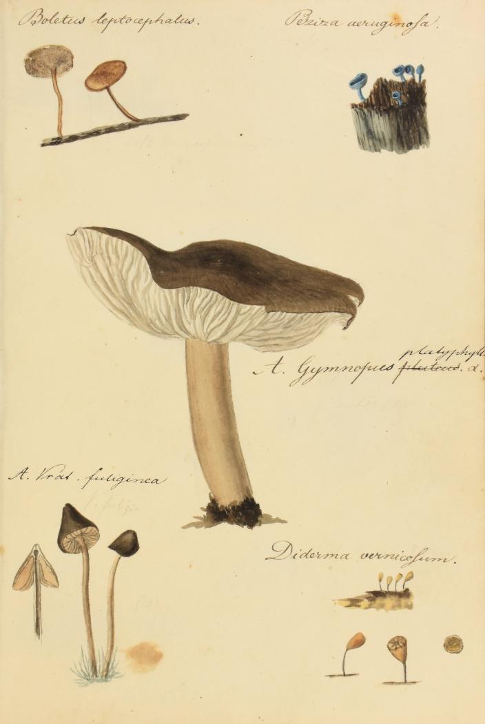
fungi illustrations

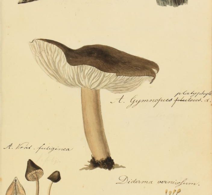
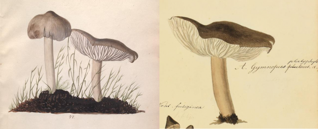
Agaricus (Gymnopus) platyphyllus. Illustration from Fungorum Nieskiensium Icones v. 2, pl. 113, fig. 77 (left) and corresponding illustration in Icones Fungorum Niskiensium, pl. 23 (right). Contributed in BHL from the Library & Archives of the Academy of Natural Sciences of Drexel University and Botany Libraries, Farlow Reference Library of Cryptogamic Botany, Harvard University.

The Harvard Icones has been digitized in the Biodiversity Heritage Library thanks to the Botany Libraries, Farlow Reference Library of Cryptogamic Botany, Harvard University, while the 5-volume set is available in BHL thanks to the Library & Archives of the Academy of Natural Sciences of Drexel University and the Wilson Library of the University of North Carolina at Chapel Hill. The Conspectus has been digitized in BHL by the LuEsther T. Mertz Library of The New York Botanical Garden.

The history of these watercolor volumes and their relationship to the Conspectus was recently examined in-depth within the article “New Light on the Mycological Work of Lewis David von Schweinitz” (2018) by Jason M. Karakehian, William R. Burk and Donald H. Pfister, published in IMA Fungus: 9(1). The article presents research made possible in-part thanks to the digital copies available in BHL.

As a sketchbook, the Harvard Icones provides a unique window into the development of Schweinitz’s research on the fungal species presented in the Conspectus. The 249 watercolor figures in the the Harvard Icones depict specimens he collected and painted during his time attending a Moravian seminary in Niesky. Most figures are labeled with a binomial in ink, although edits and annotations to the names throughout the work, as well as notes on the quality of the illustrated specimen, varieties, and colorations, illustrate the working-manuscript nature of the volume.

fungi illustration

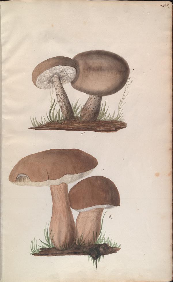
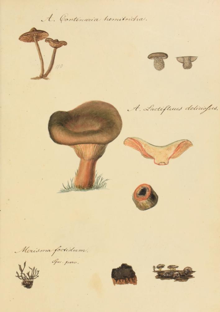
Plate 17. Merisma foetidum specimen labeled as “spec. parv.” [meager specimen]. Schweinitz, Lewis David von. Icones fungorum Niskiensium. (ca.) 1798-1802. Contributed in BHL from Botany Libraries, Farlow Reference Library of Cryptogamic Botany, Harvard University.

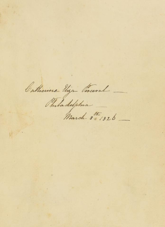
As outlined in the IMA Fungus article, the Harvard Icones has a rather convoluted history. A clue to that history can be found in the single fly-leaf bound within the volume and featuring the hand-written inscription on the recto: “Catherine Eliza Perceval — Philadelphia — March 8th 1826 —”.

fly-leaf

Fly-leaf featuring inscription for “Catherine Eliza Perceval”. Schweinitz, Lewis David von. Icones fungorum Niskiensium. (ca.) 1798-1802. Contributed in BHL from Botany Libraries, Farlow Reference Library of Cryptogamic Botany, Harvard University.

Born in Quebec in 1811, Catherine Perceval was the daughter of Anne Mary Perceval and Michael Henry Perceval. Anne had an interest in botany fueled by her association with other local female botanists, Christian Ramsay, the Countess of Dalhousie, and Harriet Campbell Sheppard. The group actively botanized together, and Anne is listed as a collector in several important botanical works of the era, including Hooker’s Flora boreali-americana ([1829]-1840) and Torrey and Gray’s A Flora of North America (1838-43).

The Harvard Icones passed into the Perceval family’s possession during a meeting in 1826 between Anne and William Darlington, a correspondent of Anne’s, at their mutual friend J. Daniel Steinhauer’s residence in Philadelphia. Schweinitz described Steinhauer as an “excellent friend…[and]…botanist”. As detailed in the IMA Fungus article, Schweinitz and Daniel Steinhauer were likely introduced by Daniel’s brother, Henry Steinhauer, who was also a friend of Schweinitz’s from his time at the Moravian seminary in Germany.

Henry had attempted to have copies made of the figures in Schweinitz’s 5-volume watercolor set, and towards this end, Schweinitz had given Henry his “Icones Fungorum Niesk’m” shortly prior to January 1818. The watercolor volume held in the Michigan Herbarium Library is the result of these endeavors. Karakehian et al. postulate that as part of this exchange the Harvard Icones may have ended up in Henry’s possession and passed to Daniel Steinhauer upon Henry’s death, after which it was passed along to Anne or directly to her daughter, Catherine (whose name is found in the Icones inscription).

fungi illustration

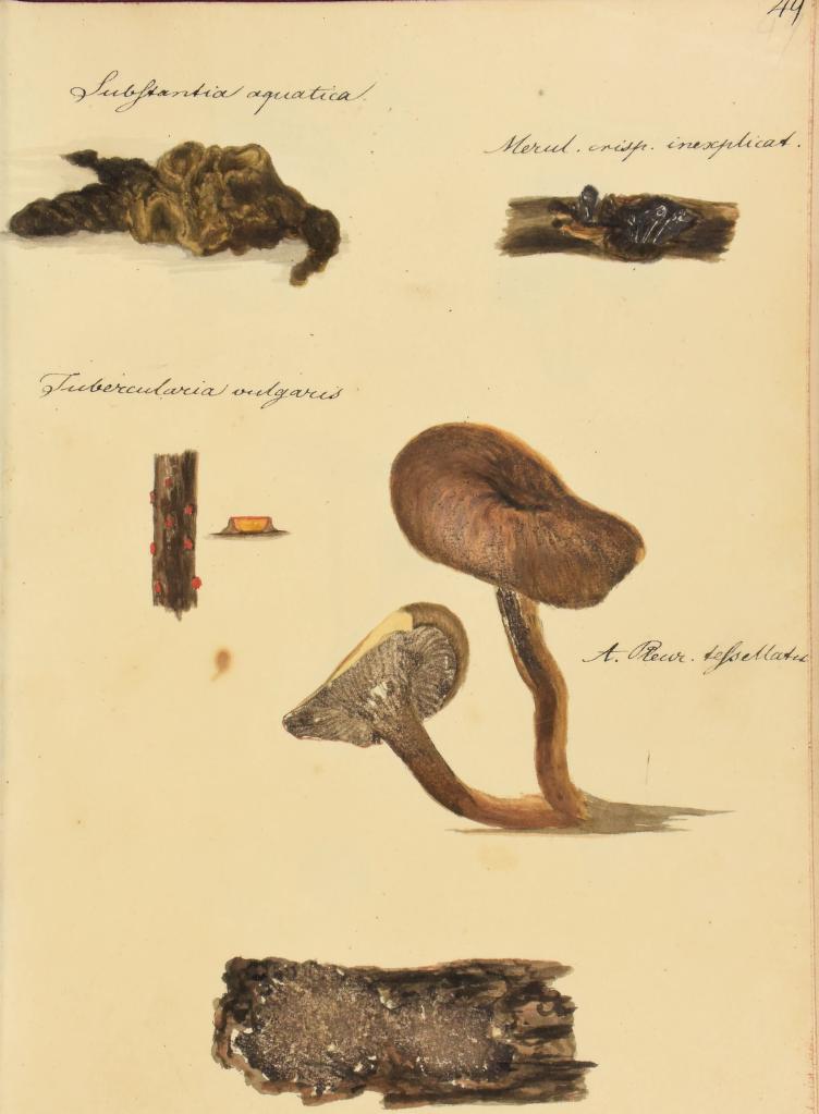
Plate 49. Merulius crispus labeled as “inexplicat[us]”, reflecting doubt as to whether the specimen was a fungus. Similarly, label of “Substantia aquatica” [watery substance]. Schweinitz, Lewis David von. Icones fungorum Niskiensium. (ca.) 1798-1802. Contributed in BHL from Botany Libraries, Farlow Reference Library of Cryptogamic Botany, Harvard University.

The volume later passed from Catherine to her son Francis Denys and eventually ended up in the possession of antiquarian book dealers and scholars William and Marianne Salloch, after whose death it was sold to a Bungay book dealer at a Christie’s auction in 1991, advertised as “Original American Watercolors” by Catherine Eliza Perceval.

Microbiologist and mycophile Elio Schaechter eventually purchased the volume from the book dealer and showed it to Donald Pfister at the Farlow Herbarium, whose research ultimately allowed him to attribute the work to Schweinitz. He also generated a manuscript index to the illustrations, which has also been digitized in BHL along with the Harvard Icones itself.

In the early 2000s, David Hewitt of the Farlow Herbarium was the first to propose that the Harvard Icones served Schweinitz as a sketchbook after comparing it to the four volumes of watercolors held at the Academy of Natural Sciences. Karakehian et al. support this conclusion, presenting further evidence within their IMA Fungus article including the incomplete quality of the sketches, the lack of any systematic arrangement of the specimens on the plates, and the annotations reflecting edits or uncertainty about species identifications, none of which are present in the five-volume set or the Conspectus.

fungi illustrations

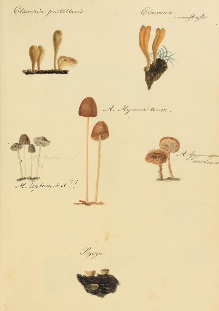
Plate 20. “Clavaria monstrosa” label indicating an anomalous specimen. Question marks indicating uncertainty over “Mycena leptocephalus” identification. Schweinitz, Lewis David von. Icones fungorum Niskiensium. (ca.) 1798-1802. Contributed in BHL from Botany Libraries, Farlow Reference Library of Cryptogamic Botany, Harvard University.

The research presented by Karakehian et al. provides novel insight into the development of a foundational mycological work. As Karakehian explains,

“These watercolor volumes are part of the knowledge-base that Schweinitz employed in his studies of American fungi. Therefore, understanding the history of these volumes is essential not only to a clearer understanding of the American mycobiota, but to an elucidation of the depth of the scientific knowledge-base at the founding of mycology in North America.”

fungi illustration

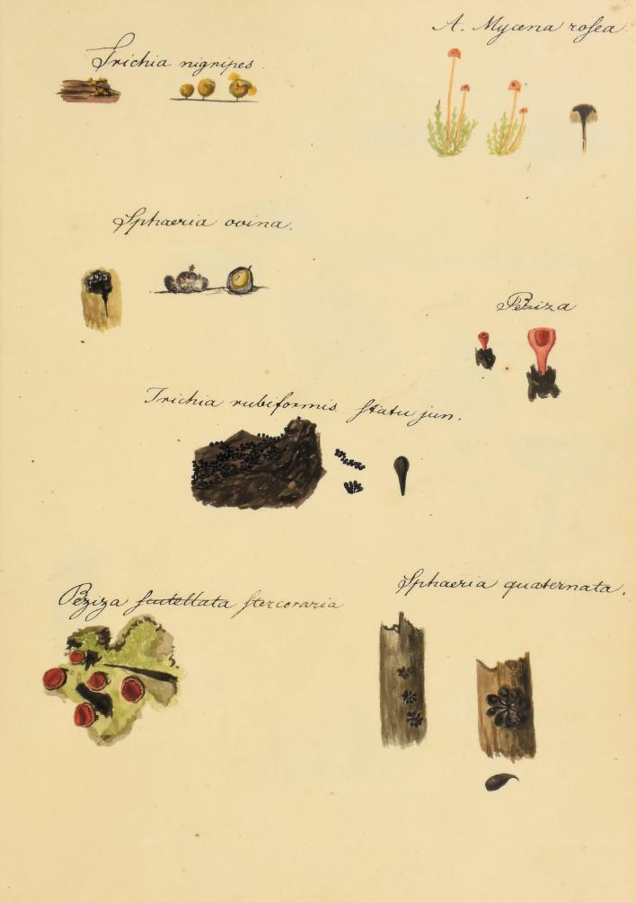
Plate 6. Trichia rubiformis labeled as “statu jun.” [immature state]. Schweinitz, Lewis David von. Icones fungorum Niskiensium. (ca.) 1798-1802. Contributed in BHL from Botany Libraries, Farlow Reference Library of Cryptogamic Botany, Harvard University.

The Harvard Icones and its related works also provide a prime example of how unprecedented research is made possible thanks to digitization initiatives like the Biodiversity Heritage Library, which allow easy reference to and comparison between volumes held in multiple institutions across the United States. As Karakehian et al. acknowledge within their paper,

“It is important to note that much of this work that resulted in this paper was made possible because archives, manuscripts and books are becoming increasingly available through digital efforts being made around the world.”

Given the unique and rare nature of the  Harvard Icones, the book was scanned at Harvard using an Internet Archive TableTop Scanner that is shared with between the Harvard Botany Libraries and the Ernst Mayr Library of the Museum of Comparative Zoology.  This mini version of an Internet Archive scribe allows Harvard librarians to scan rare or fragile materials without the wear and tear of off-site transportation. Once scanned, the Internet Archive processed the images and the final product was made accessible in the Internet Archive and BHL.  We are so grateful to the Botany Libraries, Farlow Reference Library of Cryptogamic Botany, Harvard University for all of their work to digitize the Harvard Icones in BHL and make it freely available to the world.

Similarly, we extend our sincere thanks to the LuEsther T. Mertz Library of The New York Botanical Garden, the Library & Archives of the Academy of Natural Sciences of Drexel University and the Wilson Library of the University of North Carolina at Chapel Hill, for digitizing Schweinitz’s Conspectus and other, complementary archival works, ensuring that they are also now freely available through BHL.

Take a closer look at how BHL facilitated research on these remarkable archives with our BHL User series, where Karakehian provides more insight into BHL’s impact on his work to unravel the mysteries surrounding Schweinitz’s Icones.

Post with contributions from Jason M. Karakehian (Master’s degree candidate at the Harvard Extension School), Judith A. Warnement (Librarian of Harvard University Botany Libraries), and Diane Rielinger (Digital Projects Librarian, Harvard University Herbaria & Libraries).

November 1, 2018by michelle.underhill
Blog Reel, User Stories

Mars Invaders: The Wonderful World of Microfungi

In 1897, a monumental work appeared in print for the first time. It was a story of invasion. It was a story of war. It was a story of Martians.

The story, of course, was The War of the Worlds, by H.G. Wells, which first appeared in serialized form in the UK’s Pearson’s Magazine and the US’s Cosmopolitan magazine in 1897. It was later first published in book form by William Heinemann of London in 1898. Written between 1895-97, it is one of the earliest stories centered around conflict between humans and extraterrestrials. An extremely influential work, it has never been out of print.

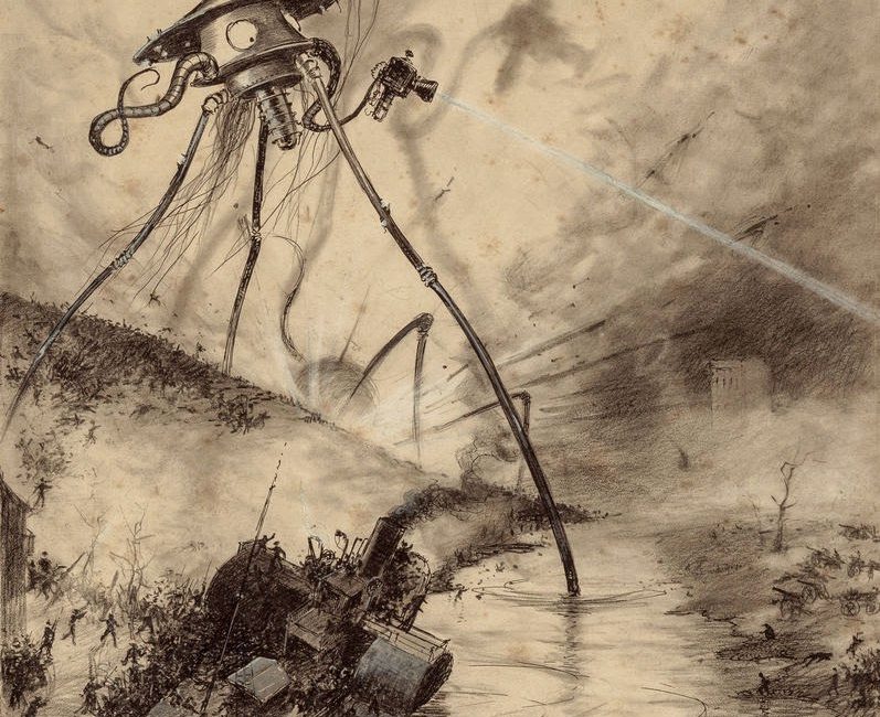
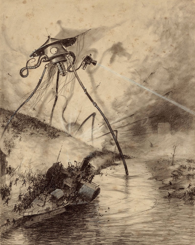
The 1906 Belgian edition of the book included drawings by Brazilian artist Henrique Alvim Corrêa. His original pencil and ink drawings, depicting Martians wreaking havoc on a devastated Earth, are in fact going up for auction today in Beverly Hills, California, and are expected to realize a sum of $500,000.

View Full Size Image
Martian Gas Cannon, from The War of the Worlds, Belgium edition, 1906. Pencil and Ink drawings by Henrique Alvim Corrêa. Image from Heritage Auctions. Image obtained from: http://www.huffingtonpost.com/2015/05/05/war-of-the-world-illustrations-auction_n_7214410.html.

Corrêa’s Martians, although largely mechanical, still have a very organic feel to them. Indeed, to the eyes of a trained expert, they bare a striking resemblance to something altogether earthly – microfungi.

Microfungi are fungi such as molds, mildews and rusts that differ from macrofungi by the absence of a large, multicellular fruiting body. Present in all terrestrial and freshwater and marine environments, microfungi grow in plants, soil, water, insects, cattle rumens, hair, and skin. Often benign, some species are also harmful, causing diseases in plants, animals and humans.

View Full Size Image
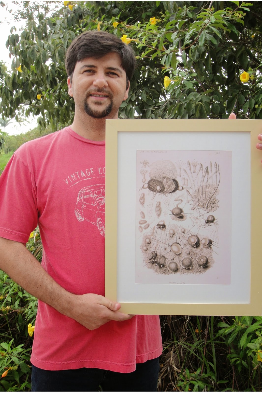
Fig. 1. Dr. Dartanhã J. Soares, plant pathologist researcher at the Brazilian Corporation for Agricultural Research (Embrapa), holding his new decoration piece which was downloaded through BHL from Tulane’s Brothers Selecta Fungorum Carpologia. Vol.1 http://biodiversitylibrary.org/page/3448217.

Dr. Dartanhã J. Soares has been studying plant pathology for twelve years. While his post graduate research focused on the taxonomy of plant pathogenic fungi, these days his work is focused on the management of fungal diseases on tropical crops at the Brazilian Agricultural Research Corporation (Embrapa). But while his current duties may deviate from an exclusive study of fungi, Dr. Soares’ true passion is still mycology.

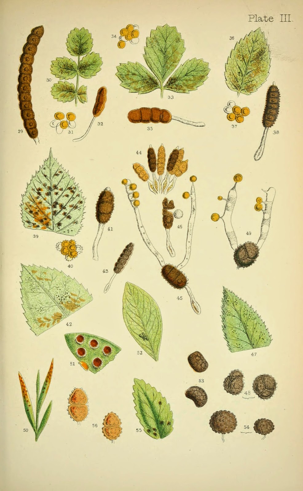
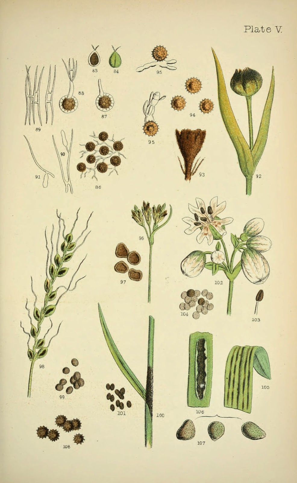
“I really love looking through the microscope and literally ‘discovering’ an amazing hidden world,” enthuses Soares. “But before that, I need to collect samples to be examined, and this is almost as exciting as looking through the microscope. I really like to go into the field and collect plant samples exhibiting the most variable range of fungal infection symptoms like those caused by rust and smut fungi. We can see some examples of symptoms and spore types in these drawings from the late nineteen century” (Figs. 2 and 3).

View Full Size Image
Fig. 2. Symptoms and spore diversity of rust fungi from Rust, smut, mildew and mould: an introduction to the study of microscopic fungi. By M.C. Cooke and illustrated by J.E. Sowerby. London, 1898. http://biodiversitylibrary.org/page/3275344.

Soares’ research on fungi, and discovery of amazing illustrations like those from Rust, smut, mildew and mould (Fig. 2 and 3), are what led him to the Biodiversity Heritage Library about seven years ago.

“I was trying to solve a puzzle about a superficial black fungus I found colonizing the leaves of Typha dominguensis,” recalls Soares. “At that time, I had just finished my Ph.D. thesis and had enough time to dig into the Internet looking for ‘obscure’ things. I eventually came across G. Arnaud’s thesis Les Astérinées, available in BHL. The book itself did not help me as much as I needed, not only because it was in French, but also because the information I was looking for was only partially mentioned. However, it revealed another hidden world which was the amazing possibility to access landmark works about fungi, of course, that I would never be able to access by any other way. However, it is important to note that I did not discover the full potential of BHL at that time. That happened later, when I ‘found’ Reginald Buller’s Researches on Fungi. At that time, [I realized], ‘Hey, I’ve already visited this website before. Maybe I can find other landmark works on it.’ And then I started to dig a little deeper into BHL.”

View Full Size Image
Fig. 3. Symptoms and spore diversity of smut fungi from Rust, smut, mildew and mould: an introduction to the study of microscopic fungi. By M.C. Cooke and illustrated by J.E. Sowerby. London, 1898. http://biodiversitylibrary.org/page/3275364.

Accessing BHL on average about once a month, Soares sees the true value of BHL through the ability to satisfy his curiosity about the natural world.

“BHL is a powerful tool and makes our lives easier,” explains Soares. “BHL is doing amazing work making such rare and wonderful ‘masterpieces’ available to everyone around the globe. Specific to my research field, BHL has a minor direct impact; sometimes it is necessary to check some old fungal description and in those cases I check BHL to see if the original work is available. However, indirectly BHL had a much bigger impact. BHL is providing me with a unique opportunity to improve my knowledge and mainly my curiosity about some classical works I had already heard about but never had the opportunity to read myself.”

While Soares most often uses BHL to download whole PDFs of books, one of his favorite features on the website is the multiple means through which he can access and download content and the breadth of materials available.

“BHL is very versatile in the ways it provides access to what I need,” lauds Soares. “Sometimes I read titles or parts of titles online, but more frequently I download the whole PDF to include the title in my personal digital library. More recently, I have also requested to download high resolution images because I was interested in having the picture framed. Additionally, at first I used BHL only for work or related topics, but now I see that there are a lot of interesting titles [outside of my direct field of study]. For example, some time ago I read the book The river of doubt: Theodore Roosevelt’s Darkest Journey by Candice Millard, and recently I saw that the book Through the Brazilian Wilderness, written by Theodore Roosevelt, is available at BHL. I have downloaded and started reading it.”

But while he loves the ability to download high resolution images from BHL, this feature also triggers a response from Soares on what he would like to see changed in BHL.

“The only thing I would change is the downloading process of high resolution images. It is a little tricky. It would be wonderful if we had the option to download high resolution images with one click, like: ‘click here to obtain this page in high resolution.'”

While we have recently released a tutorial better explaining how to download high resolution images from BHL, an easier way to provide access to these images is an item that we have added to our development wish list.

But what does all of this have to do with Martians and H.G. Wells? To understand, you have to consider Wells through the eyes of a fungi expert.

View Full Size Image
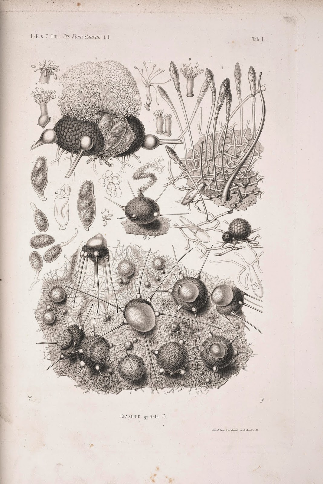
Fig. 4. Phylactinia guttata from Selecta Fungorum Carpologia. Vol.1, Tab.I, Tulasne, E.L.-R and C. 1861. http://biodiversitylibrary.org/page/34482177.

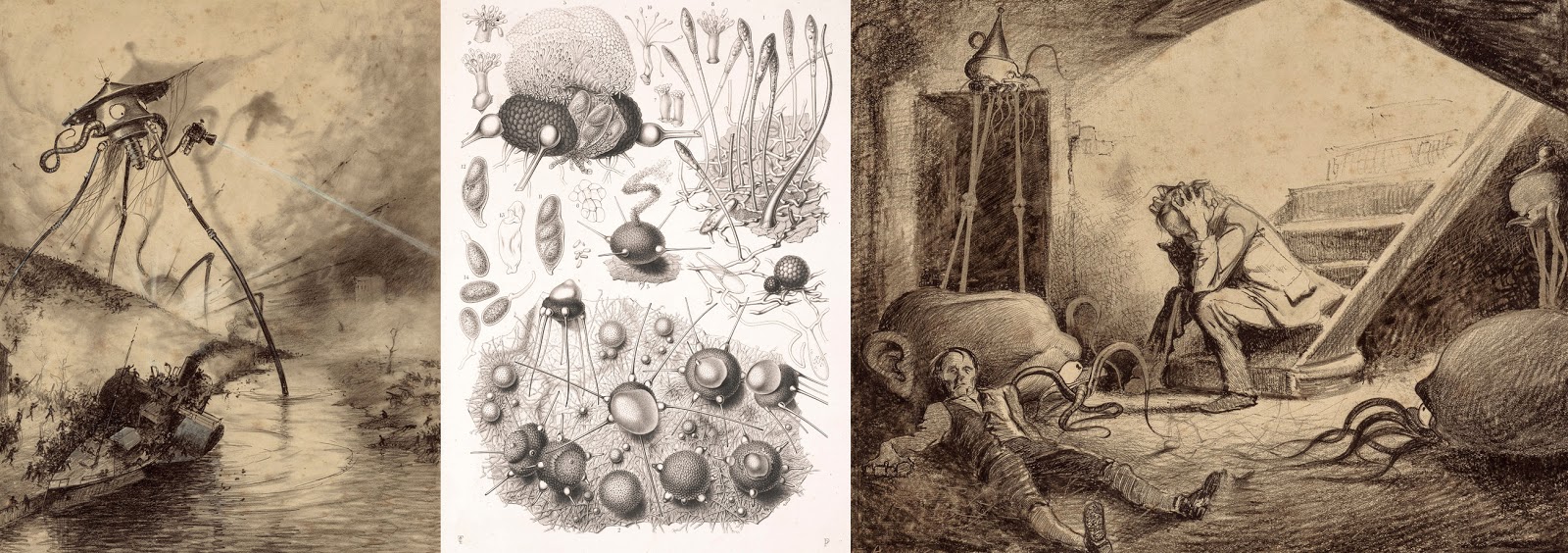
“My favorite title [in BHL] is Selecta fungorum carpologia from the Tulasne brothers,” begins Soares. “The line drawings are so amazing, so ‘real,’ and with such richness of detail that I constantly ask myself how they did that with such rudimental microscopes. If you have any doubt about this, look at Erysiphe guttata, which nowadays is called Phyllactinia guttata, in Selecta Fungorum Carpologia Vol 1, Tab.I (Fig. 4). The sexual stage, the globose bodies, of this fungus are smaller than a pin head, and even so can you see the richness of detail? Additionally, the Tulasne brothers established the pleomorphism – which is the capacity of a single organism to show distinct forms – of fungi and summarized their results in Selecta fungurom carpologia, which became a landmark [title] on fungi research. Well, at this point, you’re probably asking yourself, where are the Martians? Don’t you see them? Look more carefully at Fig. 4. Does the general aspect of the sexual bodies of P. guttata remind you of anything? No? Well, maybe you should take a second look at some of the images from The War of the Worlds. Ha-ha, probably now you are wondering if H.G. Wells got inspirations from P. guttata to describe the invading martians, aren’t you?”

View Full Size Image
Comparing Corrêa’s Martians to Tulasnes’ fungi. Left: “Martian Gas Cannon.” Right: “Death of Curate.” Both from The War of the Worlds, Belgium edition, 1906. Pencil and Ink drawings by Henrique Alvim Corrêa. Images from Heritage Auctions. Images obtained from: http://www.huffingtonpost.com/2015/05/05/war-of-the-world-illustrations-auction_n_7214410.html. Center image: Phylactinia guttata from Selecta Fungorum Carpologia. Vol.1, Tab.I, Tulasne, E.L.-R and C. 1861. http://biodiversitylibrary.org/page/3448217.

Thank you, Dr. Soares, for taking the time to explore the wonderful world of fungi and Martians with us! Do you use BHL to support your work? Want to tell us about it? Send us a message at [email protected]. 

May 14, 2015by michelle.underhill

Help Support BHL

BHL's existence depends on the financial support of its patrons. Help us keep this free resource alive!

search

About BHL

The Biodiversity Heritage Library (BHL) is the world’s largest open access digital library for biodiversity literature and archives. BHL operates as a worldwide consortium of natural history, botanical, research, and national libraries working together to digitize the natural history literature held in their collections and make it freely available for open access as part of a global “biodiversity community.”

Join Our Mailing List

Sign up to receive the latest news, content highlights, and promotions.

Subscribe Now

Subscribe to Blog via Email

Enter your email address to subscribe to this blog and receive notifications of new posts by email.

Join 319 other subscribers

Subscribe to Blog Via RSS

Subscribe to the blog RSS feed to stay up-to-date on all the latest BHL posts.

Access RSS Feed

Inspiring Discovery through Free Access to Biodiversity Knowledge.

The Biodiversity Heritage Library makes it easier than ever for you to access the information you need to study and explore life on Earth…for free, anytime, anywhere.


64+ Million Pages of
Biodiversity Literature Online.

EXPLORE

Tools and Services
to Transform Research.

EXPLORE

300,000+
Illustrations on Flickr.

EXPLORE

ABOUT | HARMFUL CONTENT | PRIVACY | SITE MAP | TERMS OF USE