Biodiversity Heritage Library - Program news and collection highlights from BHL
  • Home
  • News
  • Featured Books
    • All Featured Books
    • Book of the Month Series
    • BHL at 20
  • User Stories
  • Campaigns
    • Fossil Stories
    • Garden Stories
    • Monsters Are Real
    • Page Frights
    • Her Natural History
    • Earth Optimism 2020
  • Tech Blog
  • Visit BHL
Home
News
Featured Books
    All Featured Books
    Book of the Month Series
    BHL at 20
User Stories
Campaigns
    Fossil Stories
    Garden Stories
    Monsters Are Real
    Page Frights
    Her Natural History
    Earth Optimism 2020
Tech Blog
Visit BHL
  • Home
  • News
  • Featured Books
    • All Featured Books
    • Book of the Month Series
    • BHL at 20
  • User Stories
  • Campaigns
    • Fossil Stories
    • Garden Stories
    • Monsters Are Real
    • Page Frights
    • Her Natural History
    • Earth Optimism 2020
  • Tech Blog
  • Visit BHL
Biodiversity Heritage Library - Program news and collection highlights from BHL
Blog Reel, Featured Books

Skeletons in the Stacks: Cheselden’s Spine-Tingling Osteological Atlas

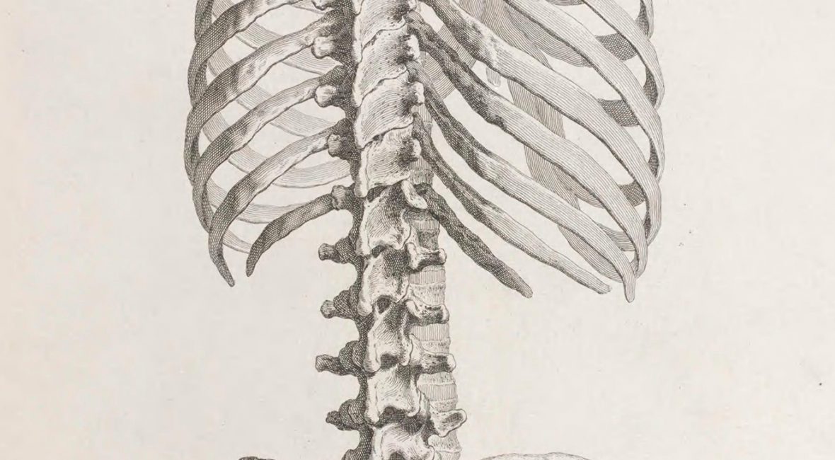
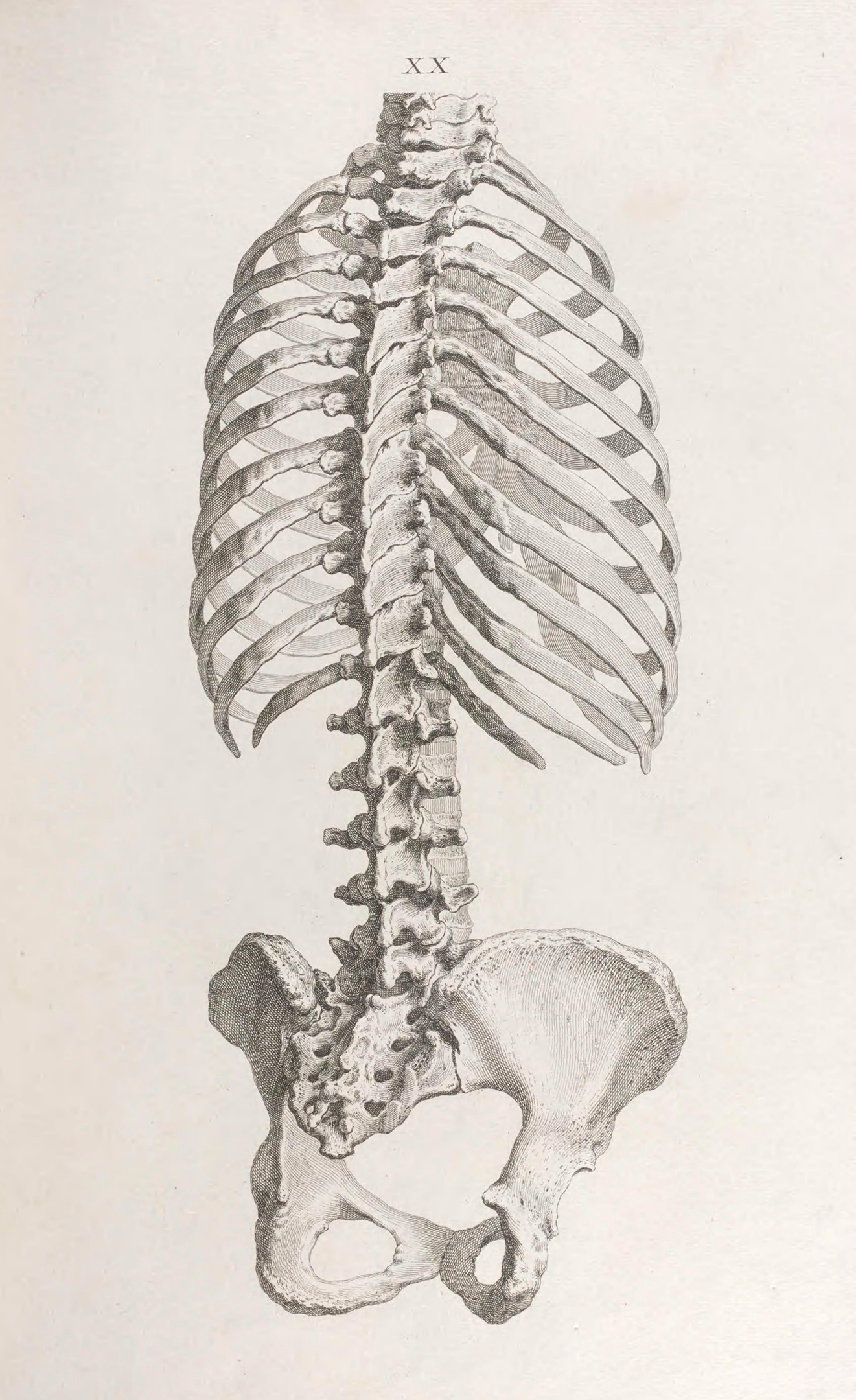
skeleton of the human torso

Plate XX. Cheselden, William. Osteographia. 1733. Contributed in BHL from the Library and Archives of the Natural History Museum, London.

Halloween is just around the corner, and the skeletons have come out of the closet. From front yards reimagined as graveyards to bone-chilling retail displays and party backdrops rattling with more than a few spare ribs, bones are on display to awaken the Halloween spirit and set a ghoulish mood to the delight of trick-or-treaters everywhere.

The modern-day Halloween is derived from the ancient Celtic festival of Samhain. With the Celtic new year celebrated on November 1, October 31 marked the end of summer and the harvest and the beginning of the cold, dark winter. Samhain, celebrated the evening of October 31, was believed to be a time when the boundary between the worlds of life and death were blurred, allowing the ghosts of the dead to return (History.com 2019).

Over time, Halloween has evolved into the costume-wearing, trick-or-treating holiday that it is today, but the season’s association with death has remained, evidenced by the proliferation of ghosts and skeletons intertwined with modern-day celebrations.

Embracing the Halloween season’s bony obsession, this month’s book of the month is one devoted to the skeleton — Osteographia (1733), which has been described as “one of the most important and beautiful books in the British anatomical tradition” (Neher 2010, 517). This work is freely available in BHL thanks to the Library and Archives of the Natural History Museum in London.
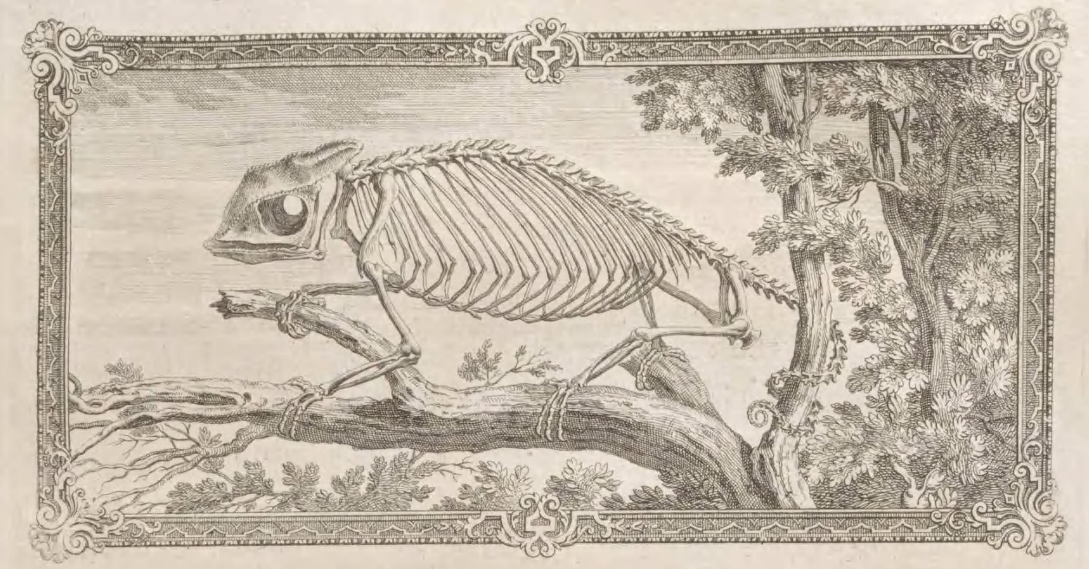
skeleton of a chameleon

Head-piece engraving for Chapter VI. Cheselden, William. Osteographia. 1733. Contributed in BHL from the Library and Archives of the Natural History Museum, London.

William Cheselden created his Osteographia with two goals in mind: to offer both the best osteological atlas and the most accurate study of the human skeleton yet produced. The book’s 56 massive plates, measuring 21 inches long and 15 inches wide, offer natural-size depictions of the separate human bones. Complete copies include two sets of plates — one set lettered with text on the verso providing descriptions of the figures, and one unlettered and blank on the verso “to shew them in their full Perfection”. Each chapter is also adorned with head and tail-piece engravings depicting various animal skeletons (Russell 1954, 38).
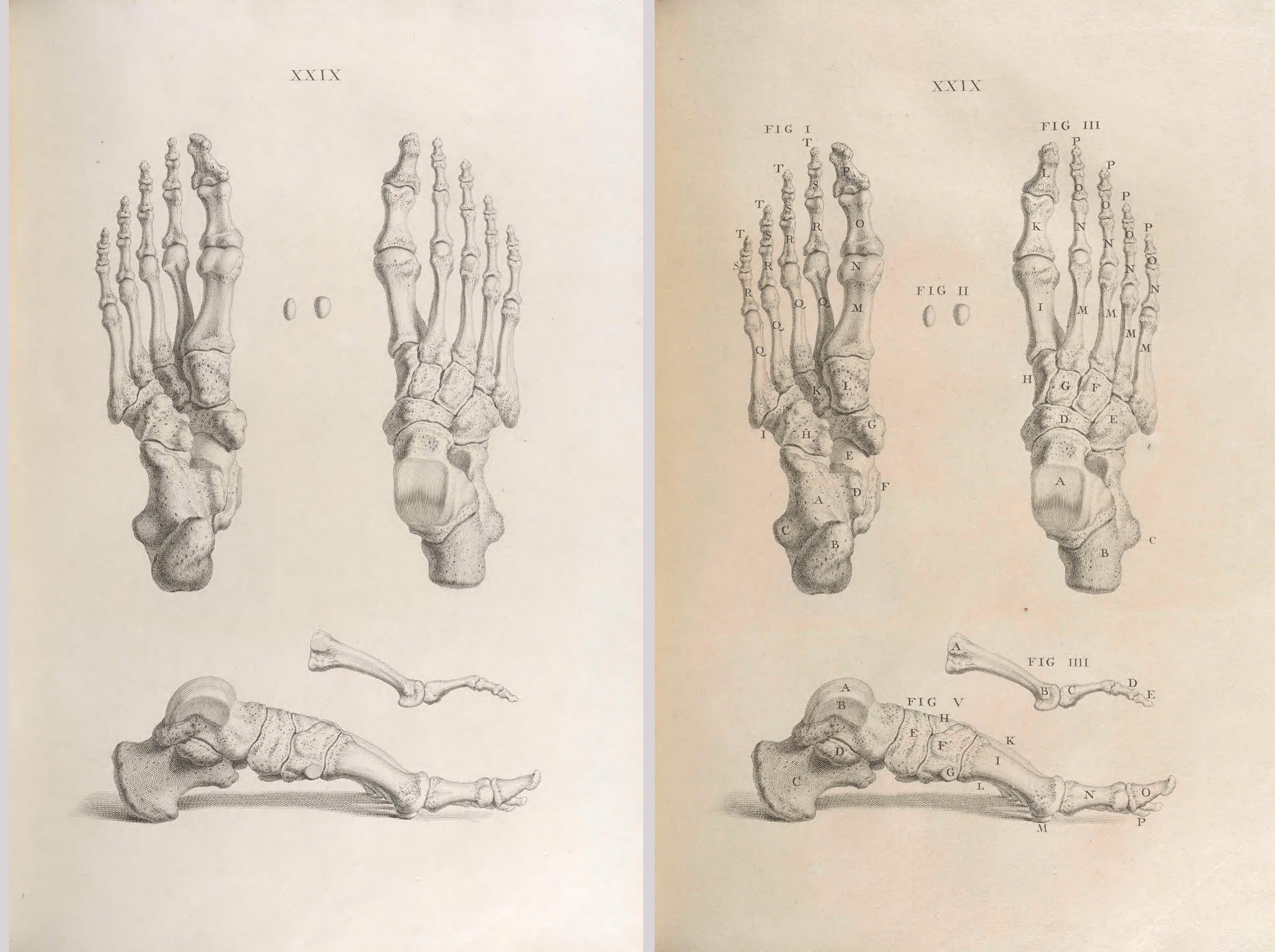
skeletons of human feet

Plate XXIX, comparison of unlettered (left) and lettered (right) plates. Complete copies include full sets of both lettered and unlettered plates. Cheselden, William. Osteographia. 1733. Contributed in BHL from the Library and Archives of the Natural History Museum, London.

Cheselden spent years engaged in the production of Osteographia. A British surgeon and teacher of anatomy and surgery, Cheselden (1688-1752) is well-known for his Anatomy of the Human Body (1713), which was used as an anatomy textbook for nearly a century. Well-respected during his time, Cheselden was one of the principal surgeons at St. Thomas’ Hospital and was appointed the surgeon to Queen Caroline in 1727 (Tikkanen 2019).
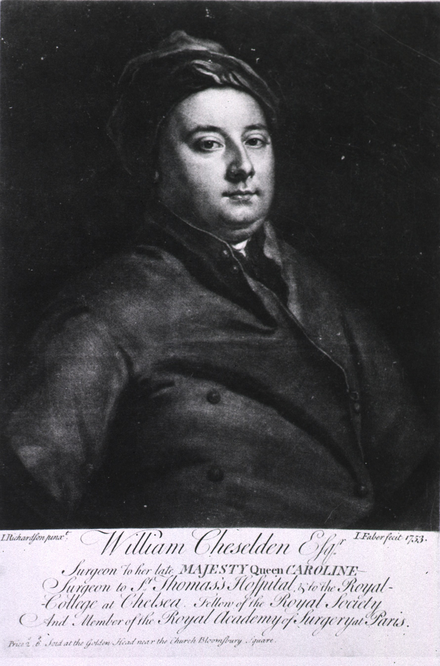
black and white portrait of a human in a coat and hat

Portrait of William Cheselden. Engraving by J. Faber after J. Richardson. 1753. Source: U.S. National Library of Medicine.

Cheselden was also a clever marketer who used the popularity of his anatomical textbook to advertise his forthcoming osteological atlas. Within the Preface to the 1726 edition of his Anatomy, Cheselden wrote that he was “much engaged” in the production of an Osteology that would provide life-size plates of the human bones. He went so far as to include an example of some of the engravings from his atlas (specifically, the maxilla and mandible figured on Plate IX of Osteographia).

Published in 1733, Cheselden limited the English edition of Osteographia to 300 copies, plus an additional 100 prints of the plates intended for a foreign edition in either Latin or French (which was never produced) (Russell 1954, 44). Following these printings, the plates were destroyed so that “the price of the book may never sink in the possession of the subscribers” (Cheselden 1733, [3]).
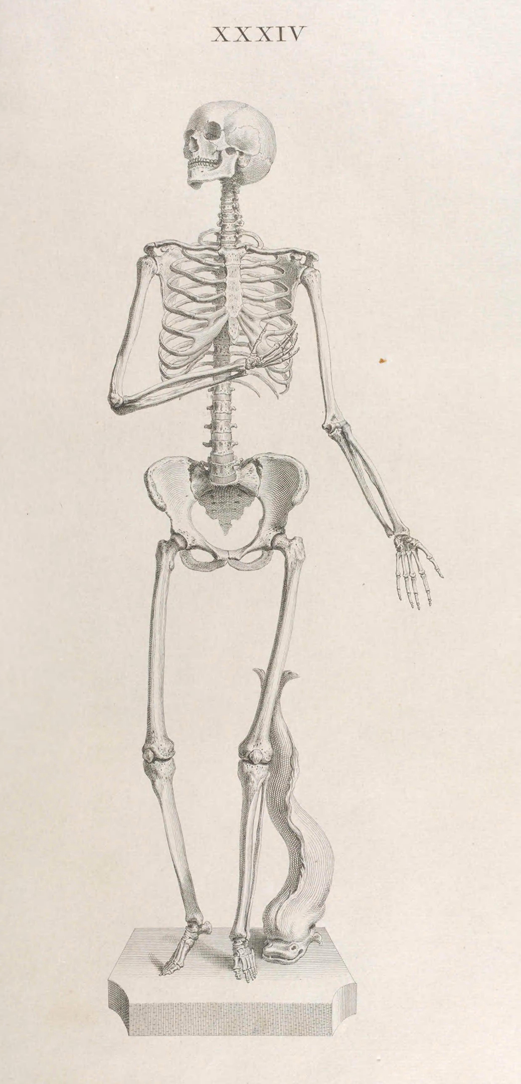
human skeleton

Plate XXXIV. Cheselden, William. Osteographia. 1733. Contributed in BHL from the Library and Archives of the Natural History Museum, London.

Cheselden’s commitment to providing the best available osteological atlas went well beyond a desire to produce an aesthetically-pleasing publication. He wanted the plates in Osteographia to serve as “working objects” for osteological studies — illustrated specimens that could be taken by the scientific community as representative. To accomplish this, accuracy of illustration was paramount (Neher 2010, 519).

To realize this ambition, Cheselden and his artists employed a camera obscura to create the initial drawings. A camera obscura is an instrument consisting of a darkened box with a convex lens or aperture that is used to project the image of an external object onto an internal screen. Cheselden is believed to be the first to use such a device for medical imagery, and the particular camera obscura used was of Cheselden’s own invention (Neher 2010, 522).
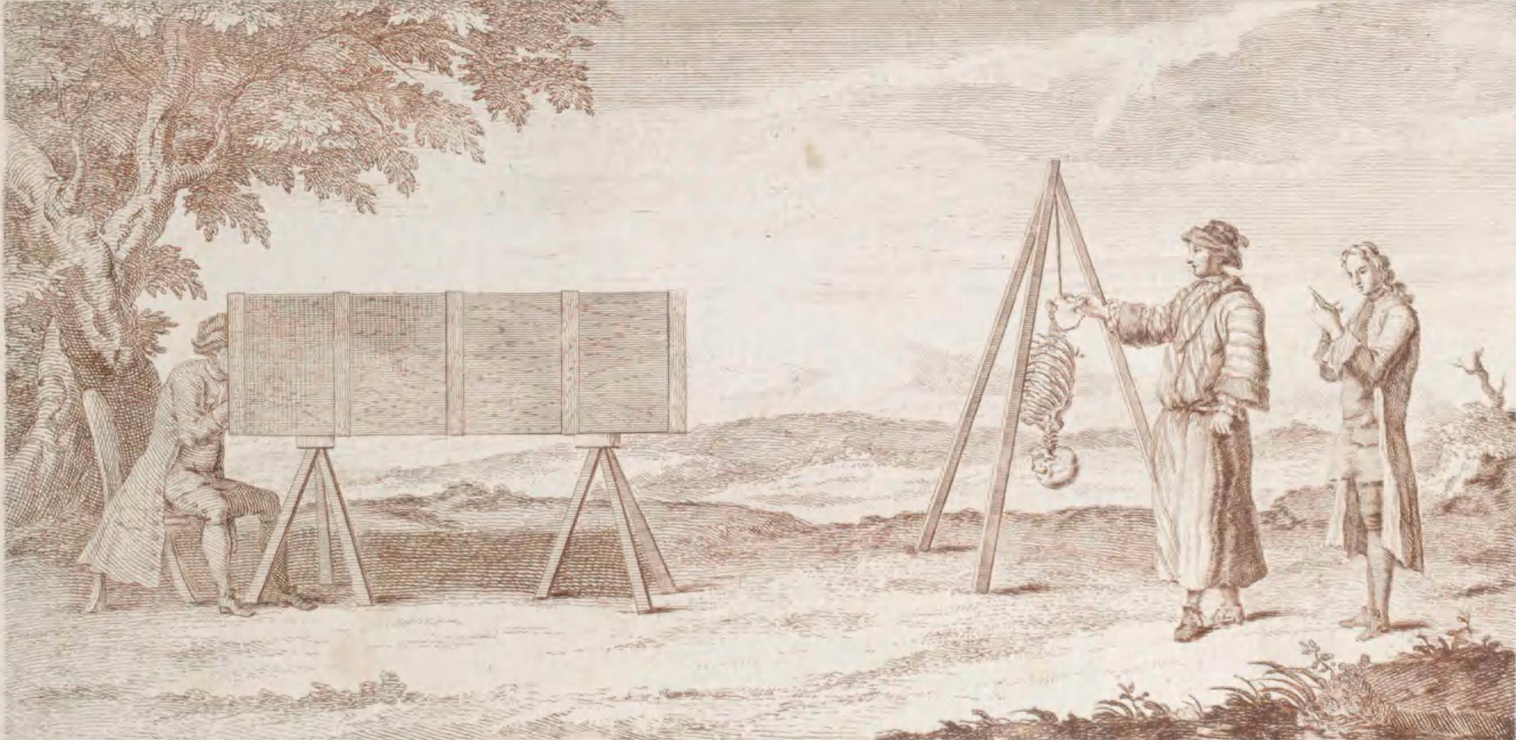
three humans using a camera obscura to illustrate a skeleton

Camera obscura figured in the vignette on the title page. Cheselden, William. Osteographia. 1733. Contributed in BHL from the Library and Archives of the Natural History Museum, London.

The camera obscura is prominently featured in the vignette on the title page of Osteographia. The two standing figures in the vignette are Cheselden’s students, John Belchier and Samuel Sharpe. The identity of the seated figure inside the camera obscura is unknown but may represent one of the artists (Neher 2010, 522).

By displaying the camera obscura so conspicuously on the title page, Cheselden was emphasizing the legitimacy and accuracy of the illustrations within Osteographia. The reader was to understand that the plates were created from actual projections of the skeletons depicted. Cheselden further listed his qualifications on the title page, including his association with the Queen and membership in several prestigious institutions, as further evidence of the credibility of the work (Neher 2010, 519).
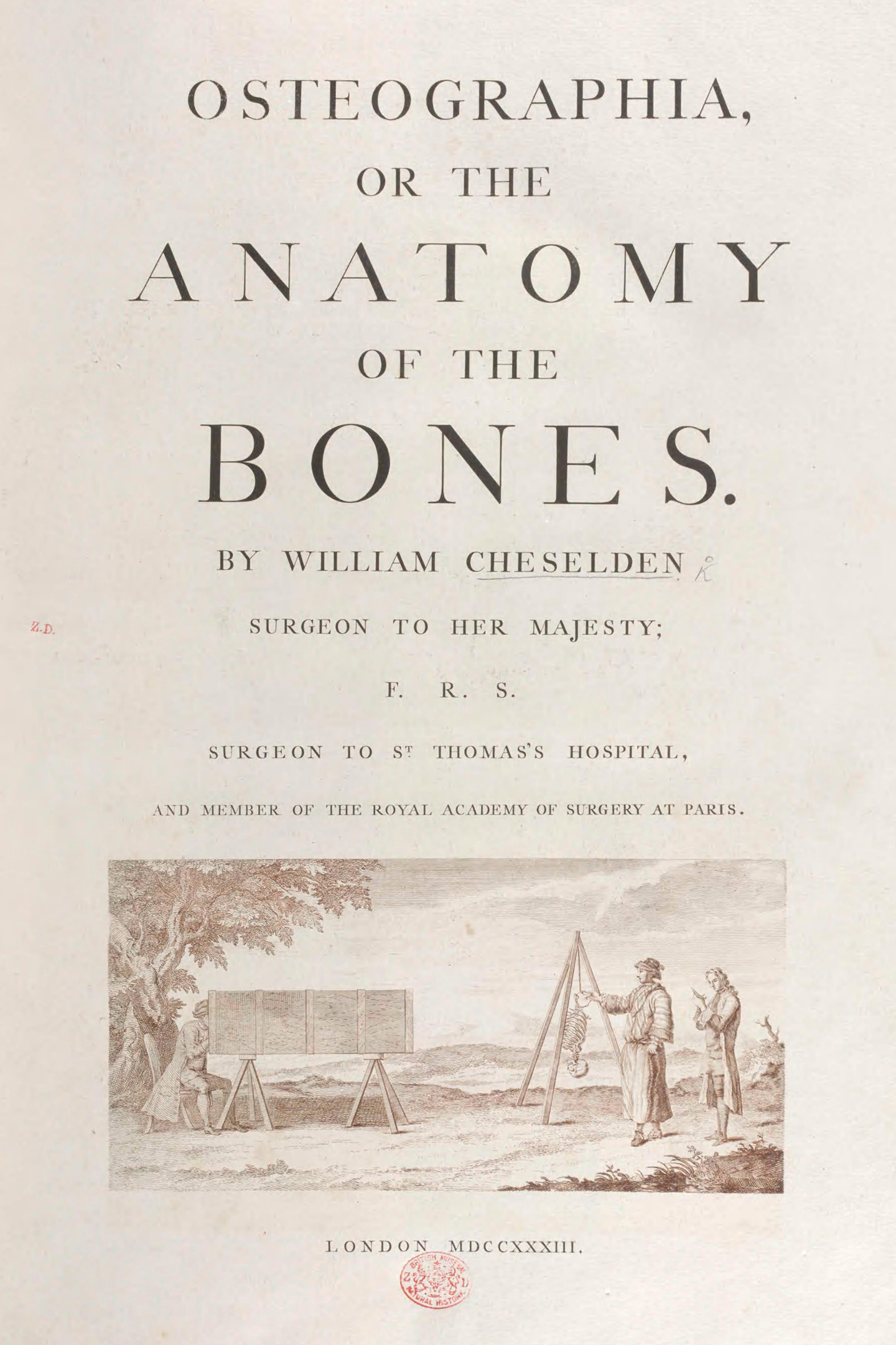
title page of Osteographia

Title page. Cheselden, William. Osteographia. 1733. Contributed in BHL from the Library and Archives of the Natural History Museum, London.

While Cheselden provided direction for the production of each image, intervening when necessary to provide corrections and alterations, the actual artists for Osteographia were Gerard van der Gucht (1695/6-1776) and Jacob Schijnvoet (1685-1733). Based on the drawings created by means of the camera obscura, the final plates were produced through a combination of etching and line engraving. Engraving, in which lines are cut into a metal plate with a burin, was used to produce smooth, dark exterior lines for each bone. Etching, in which an acid wash is used to etch an image drawn into a wax coating on a metal plate, was used for the other parts of the image (Neher 2010, 518).

Many varied issues of Osteographia exist today, as detailed by K.F. Russell in an article within Bulletin of the History of Medicine (vol. 28, no. 1, 1954). While the work was first officially published in 1733, there is a unique, 1728 trial issue in the Hunterian Library at the University of Glasgow which features 56 unlettered plates but no text or letterpress.

In addition to the “typical” 1733 issue, there is also a variant of the 1733 issue in which the title vignette and illustration of a broken column on the final page are printed in red. Additionally, this version presents the engraving of the Royal Arms on the verso of the title page, rather than on a separate page as in the “typical” 1733 issue. Russell postulates that this variant may represent a trial issue printed earlier than the “typical” 1733 issue. The copy of this work available in BHL from the Natural History Museum in London is this 1733 variant issue.
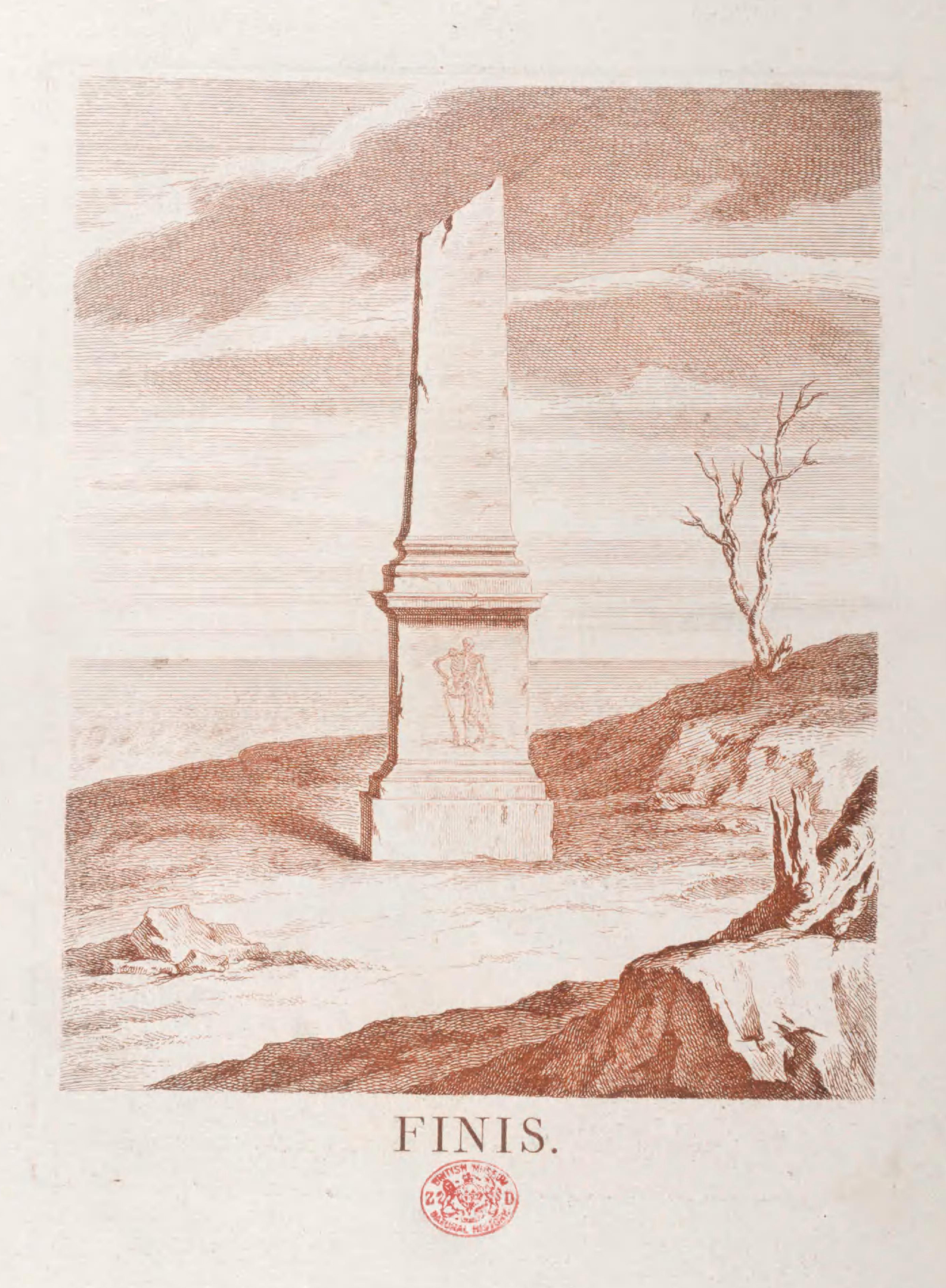
broken column with finis written below it

The broken column at the end of this copy is printed in red. This indicates that this copy is a 1733 variant issue. Cheselden, William. Osteographia. 1733. Contributed in BHL from the Library and Archives of the Natural History Museum, London.

Finally, there are several undated versions of the work which likely represent later editions made from sets of the unlettered plates broken up from the original whole and sold as separate, undated volumes in an effort to recoup some of the costs of publication.

Unfortunately, Cheselden did have need to recoup costs from the production of Osteographia. While widely praised, the work was not a commercial success. Of the 300 English copies printed, only 97 were sold by subscription. Of the remaining 203 copies, Cheselden disbound 83 of them, offering sets of the frontispieces and head pieces at 10 shillings each and sets of the 56 plates — which he advertised as “proper ornaments for a surgery” — for 30 shillings each (Russell 1954, 38-44).

Though it may not have been a commercial success, Osteographia was and is still celebrated for its beautiful and accurate illustrations, representing an important contribution to the history of medicine.

So make no bones about it — for those seeking a good skeleton or two this Halloween, Osteographia delivers in spine-tingling spades.
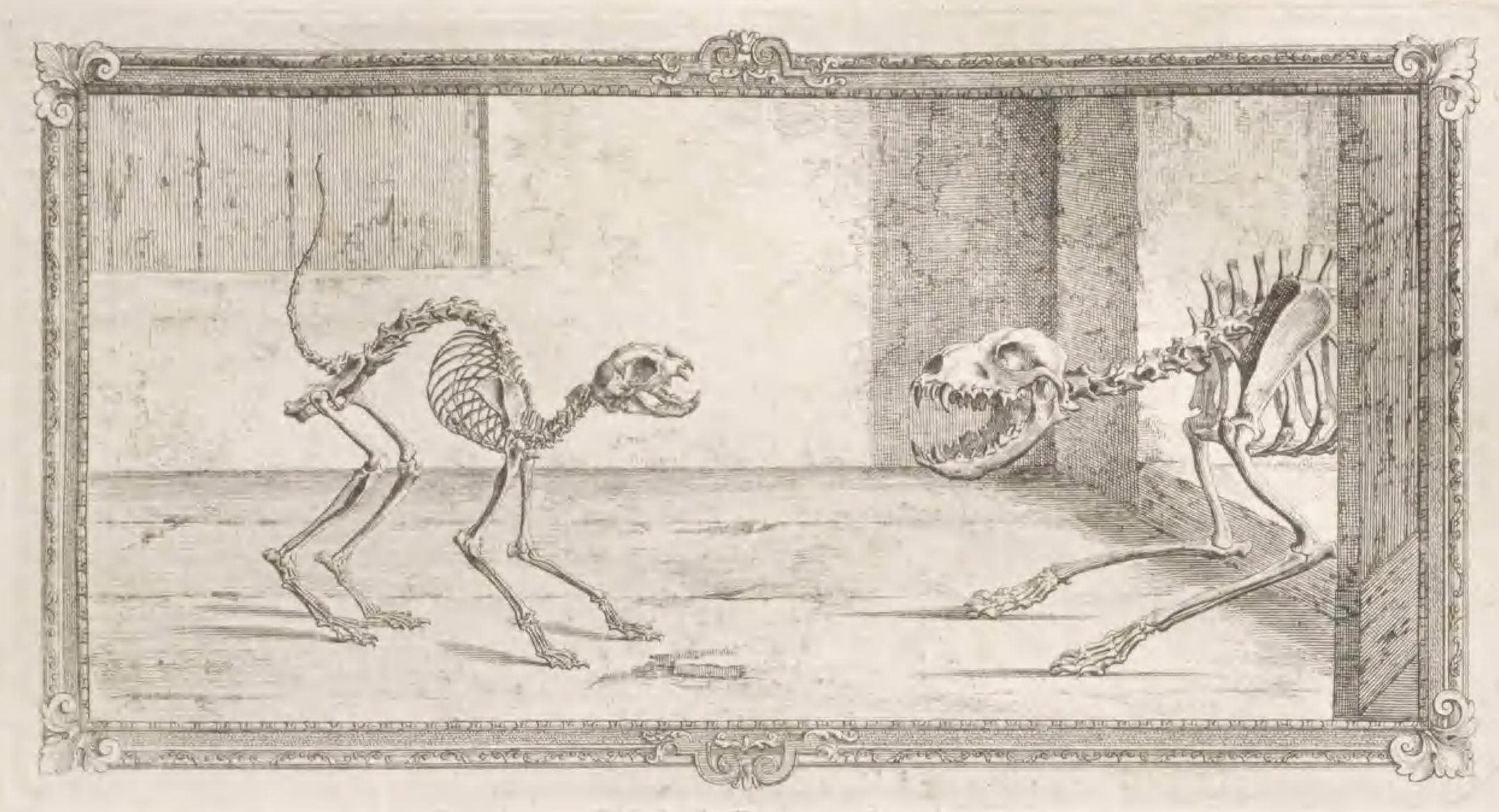
skeletons of a cat and a dog confronting each other

Head-piece engraving for Chapter II. Cheselden, William. Osteographia. 1733. Contributed in BHL from the Library and Archives of the Natural History Museum, London.

References

Cheselden, William. 1733. Osteographia. London: W Boyer.

History.com Editors. 2019. “Halloween 2019.” History.com. September 13. Accessed on October 1, 2019. https://www.history.com/topics/halloween/history-of-halloween.

Neher, Allister. 2010. “The Truth about Our Bones: William Cheselden’s Osteographia.” Medical History 54: 517-528.

Russell, K.F. 1954. “The Osteographia of William Cheselden.” Bulletin of the History of Medicine 28(1): 32-49.

Tikkanen, Amy. 2019. “William Cheselden.” Encyclopedia Britannica. April 6. Accessed on September 30, 2019. https://www.britannica.com/biography/William-Cheselden.

October 24, 2019by michelle.underhill
Blog Reel, Featured Books

John Forbes Royle: Materia Medica and Economic Botany

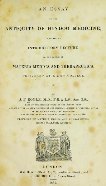
As part of the Expanding Access to Biodiversity Literature project, an interesting title was added to BHL from Yale University’s Harvey Cushing/John Hay Whitney Medical Library and the online Medical Heritage Library: An essay on the antiquity of Hindoo medicine, including an introductory lecture to the course of materia medica and therapeutics, delivered at King’s College, by physician-botanist John Forbes Royle.

Title page of An essay on the antiquity of Hindoo medicine, including an introductory lecture to the course of materia medica and therapeutics, delivered at King’s College, by Royle, J. Forbes (John Forbes), 1799-1858. London, Allen, 1837. Digitized by Yale University via the Medical Heritage Library. http://s.si.edu/2mtstTx.

This work from 1837 documents the materia medica (pharmacology) of India, and explores the historical exchange of medicinal knowledge between cultural groups of India, Arabia, Persia, Greece and China. Including details in botany, ecology, minerology and astronomy, it’s an intriguing interdisciplinary resource that can also be read for insights on its Western author and this period in time.

John Forbes Royle (1798-1858) traded his plan to join the British army for an unexpected interest in natural history. He was born at Kanpur, India, and would return to India after attending Edinburgh High School and the East India Company’s military academy at Addiscombe. Inspired by the mentorship of physician Anthony Todd Thomson, Royle chose to pursue medicine as a means to further his study of botany. He became an assistant surgeon with the East India Company, and in the following years worked at several locations across northern India, where he studied medicines from bazaars, employed collectors to amass a collection of economic plants, and became superintendent of the garden at Saharanpur. He earned the titles of MD in 1833 and Professor of Materia Medica at King’s College, London, in 1836.

An excerpt from Essay on the antiquity of Hindoo medicine demonstrates Royle’s interconnected thinking on medicine:

“There are, however, two branches of this extensive science [botany], respecting which I am desirous of making a few observations; one is the connexion between the Structure and Natural affinities of plants, and their Physical and Medical properties; and the other is the Geographical distribution, especially as connected with Climate. Both are important subjects, whether we consider them in a scientific or a practical point of view. The one teaches us the laws which influence the distribution of plants; points out the countries and climates which different families affect; and gives us principles for their cultivation, either as medicines, or as objects of agriculture: the other is no less valuable in affording us innumerable indications in every part of the world, for discovering the properties of new and unknown plants, whether as fitting them for food, for medicine, or for any of the arts of life[.]” [Royle, p. 3]

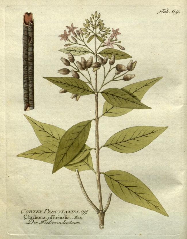
Cinchona, a South American genus. Its bark contains medicinal compounds including quinine, used to treat malaria. Royle recommended that Cinchona be grown in India. Image from BHL book: Icones plantarum medico-oeconomico-technologicarum cum earum fructus ususque descriptione. Wien:herausgegeben von Ignatz Albrecht und verlegt bey Phil. Jos. Schalbaecher, [1800]-1822. Digitized by Missouri Botanical Garden. http://s.si.edu/2iiHgMh.


Cushing/Whitney Medical Library of Yale University

The copy of Royle’s text in BHL belonged to Edward Salisbury, a Yale graduate and professor who encouraged scholarship on West and South Asia. Salisbury donated his “Oriental Library,” a collection of hundreds of rare and early printed books and manuscripts, to the college in 1870. Salisbury’s Oriental Library is foundational to what is now the third-largest collection of Islamic manuscripts in the United States. Last year, Yale celebrated Salisbury’s 175th anniversary as the first professor of Arabic and Sanskrit languages and literature in the Americas.

Essay on the antiquity of Hindoo medicine is now held at the Medical Historical Library of the Cushing/Whitney Medical Library. The library was named for Yale graduates Harvey Cushing, known as the father of neurosurgery, and John Hay Whitney, Herald Tribune editor and patron of the arts. In 1935, Cushing and two other physicians, John F. Fulton and Arnold C. Klebs, donated their personal collections of medical texts to the medical library, beginning its Medical Historical Library. Its collections include materials ranging from the 16th to 20th century, with works from Robert Boyle, Galen, William Harvey, Hippocrates, and Andreas Vesalius. Additionally, the Historical Library also holds prints, a collection of weights and measures, and presents rotating exhibits.

The Cushing/Whitney Library is also home to the Cushing Center, a room in the stacks transformed to display the medical specimens of Cushing’s Brain Tumor Registry. Cushing meticulously documented over 2,200 case studies as he pioneered the field of neurosurgery, and his registry includes specimens preserved from human surgeries and autopsies as well as notes, journal excerpts, photos and negatives.

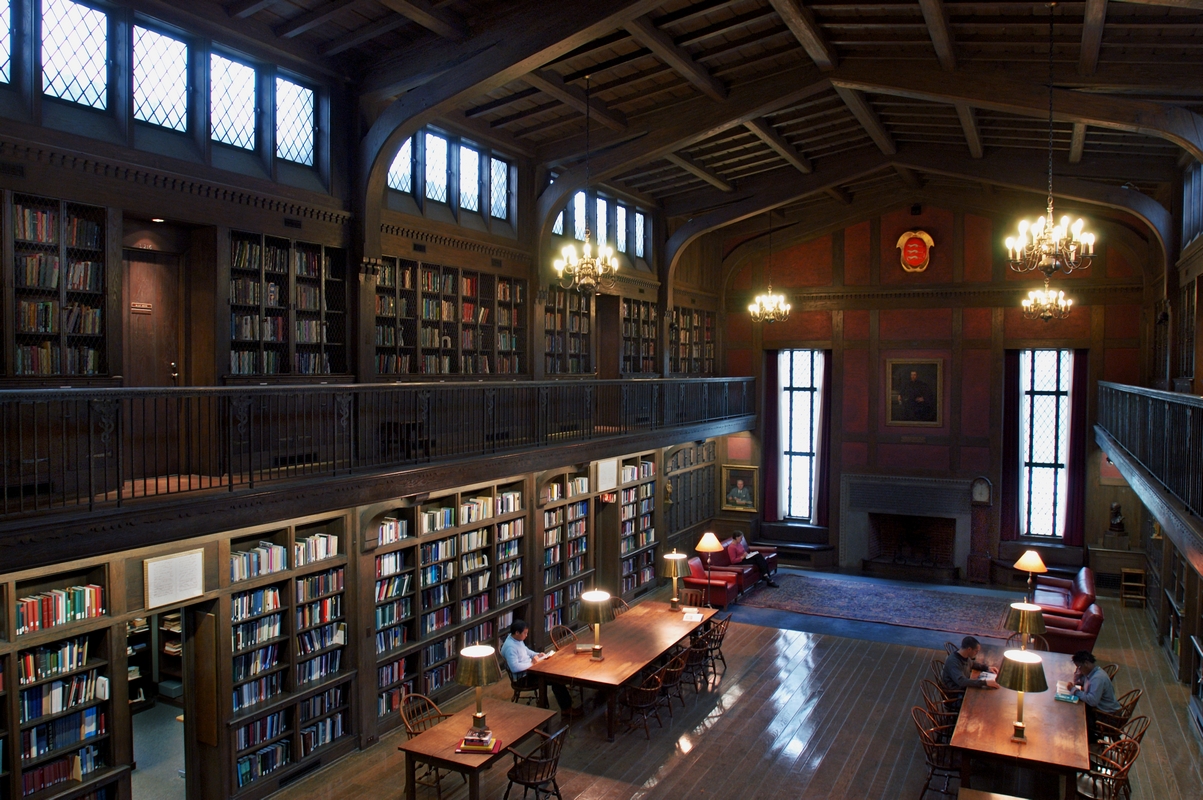
Harvey Cushing/John Hay Whitney Medical Library Historical Library. Frank Poole, 2004.


Medical Heritage Library

The Medical Heritage Library (MHL) is an online collection of materials that are free and openly accessible through the Internet Archive. Royle’s Essay on antiquity of Hindoo medicine came to BHL via the MHL.

Much like BHL, the MHL is a collaborative effort by medical libraries to promote access to resources (such as rare books, pamphlets, journals and films) that are useful across a spectrum of disciplines. The collection includes over 200,000 titles with fascinating variety – skimming the landing page, one can find herbals, medical dictionaries and papers, as well as video clips of 1960s tobacco commercials and Helen Keller’s autobiography.

The Medical Heritage Library was launched in 2010 by founders The Augustus C. Long Health Sciences Library at Columbia University and Columbia University Libraries/Information Services; The College of Physicians of Philadelphia; The Cushing/Whitney Medical Library at Yale University; The Francis A. Countway Library of Medicine at Harvard University; U.S. National Library of Medicine; and Wellcome Library in London, UK. (For more information about its content contributors, funding and support, please see MHL’s ‘About’ page.)

An essay on the antiquity of Hindoo Medicine is a welcome addition to BHL’s collection of works by John Forbes Royle, which include:

  • Illustrations of the botany and other branches of the natural history of the Himalayan Mountains: and of the flora of Cashmere. London: Wm. H. Allen, 1839. (Digitized by Missouri Botanical Garden)
  • On the culture and commerce of cotton in India and elsewhere; with an account of the experiments made by the Hon. East India Company up to the present time. London: Smith, Elder, 1851. (Digitized by the University of British Columbia Library)
  • The fibrous plants of India, fitted for cordage, clothing, and paper. With an account of the cultivation and preparation of flax, hemp, and their substitutes. London, Smith, Elder, and Co.; 1855. (Digitized by Cornell University Library)
  • A manual of materia medica and therapeutics including the preparations of the pharmacopoeias of London, Edinburgh, and Dublin, with other approved medicines. London: John Churchill, MDCCCLVI [1856]. (Digitized by King’s College London, Foyle Special Collections Library)

Thanks to Melissa Grafe, Ph.D, Head of the Medical Historical Library and John R. Bumstead Librarian for Medical History, who shared how this volume came to the Medical Historical Library.

Sources

  • About (n.d.) Retrieved from http://archive.org/details/medicalheritagelibrary&tab;=about.
  • About (n.d.) Retrieved from http://www.medicalheritage.org/about/.
  • About (n.d) Retrieved from https://salisbury175.yale.edu/about.
  • Harvey Cushing and the Cushing Center (n.d.) Retrieved from http://library.medicine.yale.edu/cushingcenter/history.
  • History of the Library (n.d.) Retrieved from http://library.medicine.yale.edu/about/history.
  • Royle, John Forbes (1837). London, Allen. Retrieved from https://www.biodiversitylibrary.org/item/199214.
  • Woodward, B. B. “Royle, John Forbes (1798–1858).” Rev. Mark Harrison. Oxford Dictionary of National Biography. Ed. H. C. G. Matthew and Brian Harrison. Oxford: OUP, 2004. Online ed. Ed. David Cannadine. May 2010. Retrieved from http://www.oxforddnb.com/view/article/24239.
November 16, 2017by ulib-libraryjobs
Blog Reel, Featured Books

Jesús Sánchez’s Mexican Medical Zoology

Sánchez, Jesús. Datos para la zoología médica mexicana: arácnidos é insectos. México: Oficina Tipográfica de la Secretaría de Fomento, 1893. viii, 189, iv pages, 9 leaves of plates : illustrations ; 23 cm.

View Full Size Image
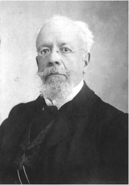
Parra, Alfonso. Atlas histórico de la Escuela Nacional Preparatoria: desde su fundación hasta los momentos de celebrarse el centenario de la proclamación de la Independencia, septiembre 15 de 1910 / México : [Escuela Nacional Preparatoria], 1910. 1 vol.

With the single purpose of serving the country and with the belief that scientific study should be used with practical ends, Doctor Jesús Sánchez elaborated his essay on Mexican Medical Zoology (Datos para la zoología médica mexicana), where he acknowledges “the value of studying the animal kingdom, within which exists a vast variety that can offer both benefits and diseases”.

Equally, he states that it “is not only important to study the organisms living inside our own bodies, it is also necessary to know those whose venoms, when inoculated in the human body, can cause, from very light inconveniences, to very serious symptoms and even death”. That is why the author developed an essay about this topic, identifying, describing and studying arachnids and insects from several parts of Mexico that cause damage to the human health.

Doctor Jesús Sánchez was a physician by profession, a member of the Academy of Medicine, and responsible of the professorships in Zoology and Biology at the National High School and the National School of Agriculture, where he has dedicated several years to the compilation of insects; these where included in his work of Mexican Medical Zoology, published in 1893. A second edition or reprint of this work has never been published.

View Full Size Image
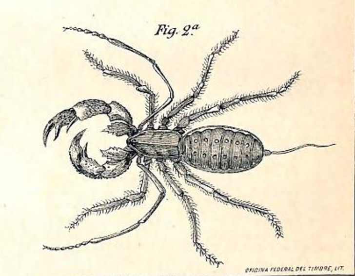
Thelyphonus giganteus. Vinagrillo. (Seg. Lucas). http://biodiversitylibrary.org/page/51139808.

During the development of this work, Doctor Sánchez exchanged knowledge, talks and experiences with the doctors and brothers Alfredo and Eugenio Dugés. Alfredo Dugés was a great illustrator who made a perfect descriptive work of the Aquatic Spider, or hydroarachnid, native of Guanajuato. Dugés directed Sánchez and recommended the research of other zoology scholars, like Fernando Altamirano, Lauro Jimenez, Antonio Peñafiel, Alfonso Herrera and Gumersindo Mendoza, among others, as well as other prominent publications from the naturalists Francisco J. Clavijero and Jose Alzate Ramirez.

Datos para la zoología médica mexicana describes, in a detailed manner, nine orders of arachnids and scorpions, as well as five orders of insects, ants and bees, using a mixture of a colloquial and technical language, explaining symptoms, cures and medicines that can be used to fight against symptoms caused by the bites or contact with insects. Within his work we can find a remarkably detailed index of every order and term, as well as illustrations for context.

View Full Size Image
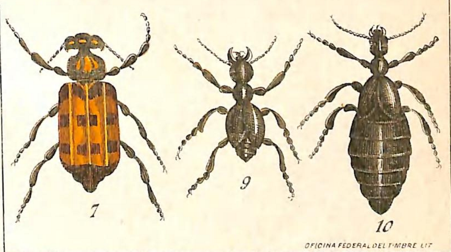
7. Horia maculata. 9. Treiodous barranci (macho). 10. Treiodous barranci (hembra). http://biodiversitylibrary.org/page/51139812.

One can appreciate nine pages that contain the illustrations of arachnids, scorpions, several insects, ants and bees, elaborated by hand with colours identical to those of the studied animals.

Being a professor of the National High School, Sánchez’s published works supported the students with knowledge and practical applications; he offered his expertise during his lectures and turned new students towards the development of science and knowledge.

View Full Size Image
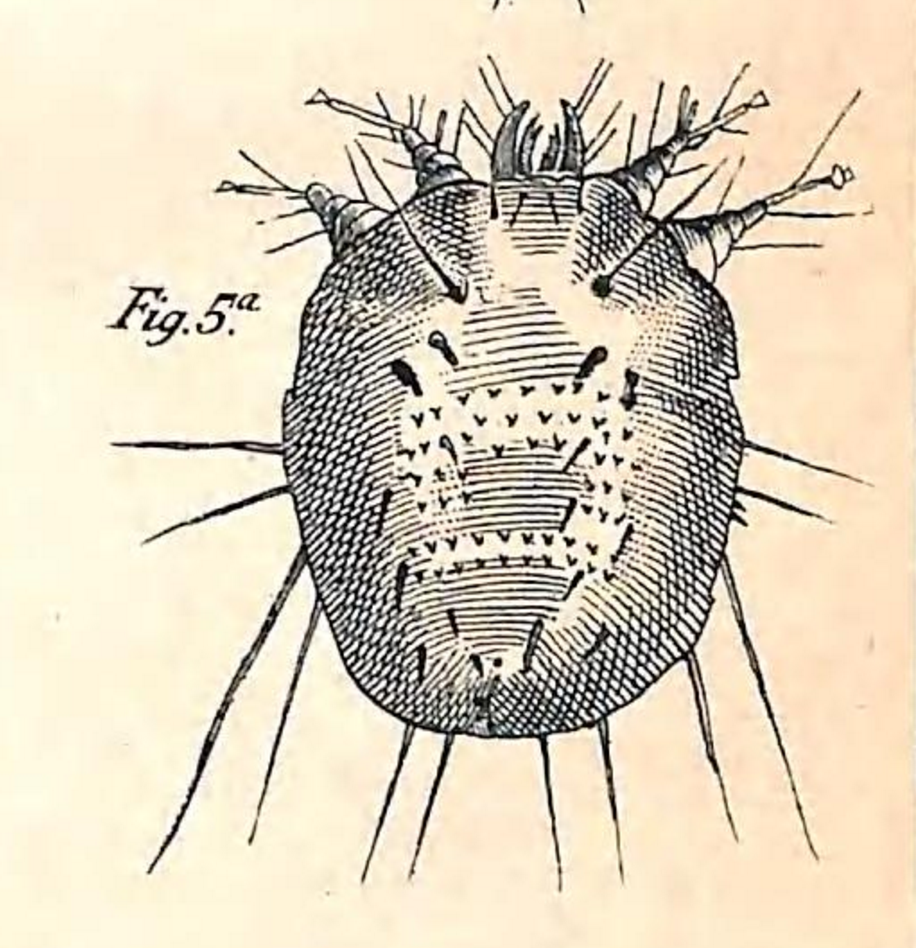
Sarcoptes scabiei (hembra). Visto por el dorso. (seg. Gudden). http://biodiversitylibrary.org/page/51139802.

Doctor Jesús Sánchez is also the author of other works related to the Mexican Medical Zoology of arachnids and insects, such as: Elementos de historia natural en forma de lecciones de cosas: Obra escrita expresamente para uso de las escuelas primarias de la nación, published in the year 1895 and available in BHL thanks to the Joint Library of Earth Sciences (Biblioteca Conjunta de Ciencias de la Tierra, Instituto de Ciencias del Mar y Limnología, UNAM), and the introduction of the work: Breve noticia de los establecimientos de instrucción dependientes de la Secretaría de Estado y del Despacho de Justicia e Instrucción Pública, edited in the year 1900.

View Full Size Image
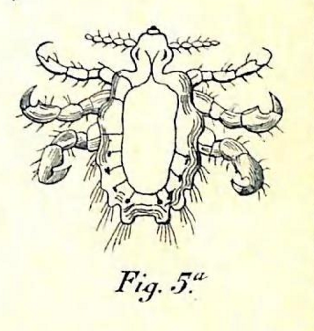
Ladilla o piojo del pubis. http://biodiversitylibrary.org/page/51139818.


September 22, 2016by ulib-libraryjobs
Blog Reel, Featured Books

Eerie Anatomy: Vesalius’ De humani corporis fabrica

This post originally published on the Smithsonian Libraries blog Unbound. See it here.

Halloween is quickly approaching and with it comes the traditional decorations of bats, pumpkins, ghosts and of course, skeletons. Back in the 1500’s, one man changed the way the medical world saw the skeletal and muscular systems of the human body. That man, Andreas Vesalius, illustrated anatomical features in his De humani corporis fabrica (On the structure of the human body) in a way never before seen. Although the pages below may seem pretty gruesome, they come from one of the most influential anatomy books of all time.

View Full Size Image
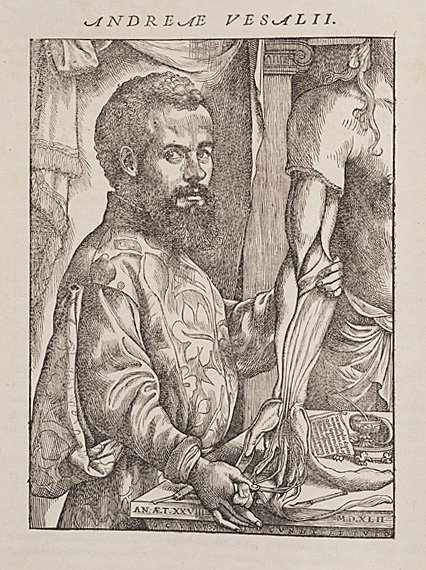
Portrait of Vesalius from De humani corporis fabrica, 1543. http://biodiversitylibrary.org/page/48714916.

Born in Brussels in 1514, Andreas Vesalius came from a family of doctors and apothecaries. When he became a physician himself and began teaching medical school in the 1530’s, most of the standard texts were based on animal dissections and the observations of an ancient Roman doctor named Galen. Vesalius took the unconventional step of performing dissections live in front of his medical students and using human bodies, in some cases those of convicted criminals.

View Full Size Image
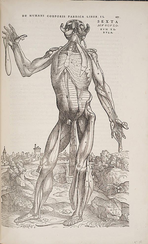
Standing figure from De humani corporis fabrica, 1543. http://biodiversitylibrary.org/page/48715058.

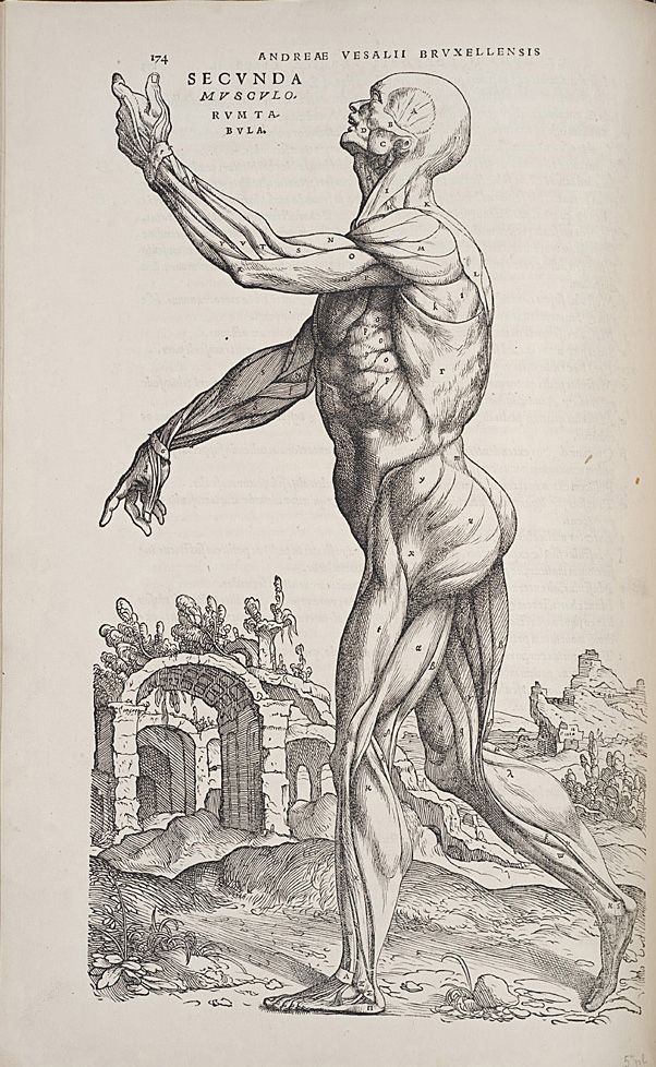
In 1543, 29 year old Vesalius published De humani corporis fabrica with elaborate wood-cut illustrations of human dissections. But these bodies weren’t just lying still, as if on a mortuary slab. They were depicted in classical contrapposto. In many instances, layers of tissue artfully fall away to reveal the muscles and ligaments which lie beneath. Despite the artistic quality, the book is considered one of the first anatomical works, accurately representing and describing various systems in the body.

Vesalius gave detailed notes to the printer, helping to assure that the descriptive text matched the appropriate figure and that the artistic style of the plates was preserved. The printer, Johannes Oporinus, felt that these notes were so important that he included them in the publication itself (see “Printer’s Note to the Reader” here: http://vesalius.northwestern.edu/books/FA.b.html). In the text of the book, Vesalius not only describes the systems of the body, but also his recommendations for obtaining skeletons, putting them together and dissecting cadavers.

View Full Size Image
Standing figure from De humani corporis fabrica, 1543. http://biodiversitylibrary.org/page/48715045.

The so-called “Muscle Men” of the book are depicted on a background that is thought to be Padua, where Vesalius taught medicine. If pieced together, the plates’ backgrounds form a full landscape. Vesalius dedicated the book to Holy Roman Emperor Charles V and later went to work as the Emperor’s personal physician.

You can learn more about Vesalius and the history of anatomy books during a live tour in the Smithsonian’s Dibner Library of the History of Science and Technology via Periscope on Thursday, October 29th at 1pm (EST). Head of Special Collections, Lilla Vekerdy, will highlight a few of the rare books, including Vesalius’, that helped transform the way that medical professions studied the human body.

View Full Size Image

If you’d like to explore more of De humani corporis fabric on your own, check out the recently-added copy from the Getty Research Institute (via the Internet Archive) in BHL.

We hope you’ll join us on Oct. 29 for the Eerie Anatomy Periscope. Make no bones about it, it will be a ghoulishly good time!

October 28, 2015by ulib-libraryjobs

Help Support BHL

BHL's existence depends on the financial support of its patrons. Help us keep this free resource alive!

search

About BHL

The Biodiversity Heritage Library (BHL) is the world’s largest open access digital library for biodiversity literature and archives. BHL operates as a worldwide consortium of natural history, botanical, research, and national libraries working together to digitize the natural history literature held in their collections and make it freely available for open access as part of a global “biodiversity community.”

Join Our Mailing List

Sign up to receive the latest news, content highlights, and promotions.

Subscribe Now

Subscribe to Blog via Email

Enter your email address to subscribe to this blog and receive notifications of new posts by email.

Join 319 other subscribers

Subscribe to Blog Via RSS

Subscribe to the blog RSS feed to stay up-to-date on all the latest BHL posts.

Access RSS Feed

Inspiring Discovery through Free Access to Biodiversity Knowledge.

The Biodiversity Heritage Library makes it easier than ever for you to access the information you need to study and explore life on Earth…for free, anytime, anywhere.


64+ Million Pages of
Biodiversity Literature Online.

EXPLORE

Tools and Services
to Transform Research.

EXPLORE

300,000+
Illustrations on Flickr.

EXPLORE

ABOUT | HARMFUL CONTENT | PRIVACY | SITE MAP | TERMS OF USE