Biodiversity Heritage Library - Program news and collection highlights from BHL
  • Home
  • News
  • Featured Books
    • All Featured Books
    • Book of the Month Series
    • BHL at 20
  • User Stories
  • Campaigns
    • Fossil Stories
    • Garden Stories
    • Monsters Are Real
    • Page Frights
    • Her Natural History
    • Earth Optimism 2020
  • Tech Blog
  • Visit BHL
Home
News
Featured Books
    All Featured Books
    Book of the Month Series
    BHL at 20
User Stories
Campaigns
    Fossil Stories
    Garden Stories
    Monsters Are Real
    Page Frights
    Her Natural History
    Earth Optimism 2020
Tech Blog
Visit BHL
  • Home
  • News
  • Featured Books
    • All Featured Books
    • Book of the Month Series
    • BHL at 20
  • User Stories
  • Campaigns
    • Fossil Stories
    • Garden Stories
    • Monsters Are Real
    • Page Frights
    • Her Natural History
    • Earth Optimism 2020
  • Tech Blog
  • Visit BHL
Biodiversity Heritage Library - Program news and collection highlights from BHL
Blog Reel, Featured Books

Eerie Anatomy: Vesalius’ De humani corporis fabrica

This post originally published on the Smithsonian Libraries blog Unbound. See it here.

Halloween is quickly approaching and with it comes the traditional decorations of bats, pumpkins, ghosts and of course, skeletons. Back in the 1500’s, one man changed the way the medical world saw the skeletal and muscular systems of the human body. That man, Andreas Vesalius, illustrated anatomical features in his De humani corporis fabrica (On the structure of the human body) in a way never before seen. Although the pages below may seem pretty gruesome, they come from one of the most influential anatomy books of all time.

View Full Size Image
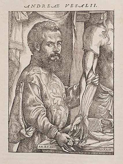
Portrait of Vesalius from De humani corporis fabrica, 1543. http://biodiversitylibrary.org/page/48714916.

Born in Brussels in 1514, Andreas Vesalius came from a family of doctors and apothecaries. When he became a physician himself and began teaching medical school in the 1530’s, most of the standard texts were based on animal dissections and the observations of an ancient Roman doctor named Galen. Vesalius took the unconventional step of performing dissections live in front of his medical students and using human bodies, in some cases those of convicted criminals.

View Full Size Image
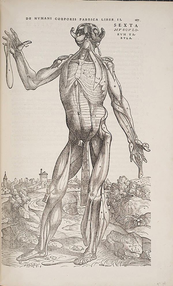
Standing figure from De humani corporis fabrica, 1543. http://biodiversitylibrary.org/page/48715058.

In 1543, 29 year old Vesalius published De humani corporis fabrica with elaborate wood-cut illustrations of human dissections. But these bodies weren’t just lying still, as if on a mortuary slab. They were depicted in classical contrapposto. In many instances, layers of tissue artfully fall away to reveal the muscles and ligaments which lie beneath. Despite the artistic quality, the book is considered one of the first anatomical works, accurately representing and describing various systems in the body.

Vesalius gave detailed notes to the printer, helping to assure that the descriptive text matched the appropriate figure and that the artistic style of the plates was preserved. The printer, Johannes Oporinus, felt that these notes were so important that he included them in the publication itself (see “Printer’s Note to the Reader” here: http://vesalius.northwestern.edu/books/FA.b.html). In the text of the book, Vesalius not only describes the systems of the body, but also his recommendations for obtaining skeletons, putting them together and dissecting cadavers.

View Full Size Image
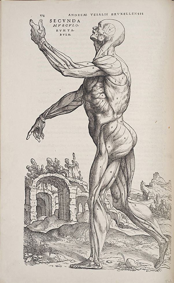
Standing figure from De humani corporis fabrica, 1543. http://biodiversitylibrary.org/page/48715045.

The so-called “Muscle Men” of the book are depicted on a background that is thought to be Padua, where Vesalius taught medicine. If pieced together, the plates’ backgrounds form a full landscape. Vesalius dedicated the book to Holy Roman Emperor Charles V and later went to work as the Emperor’s personal physician.

You can learn more about Vesalius and the history of anatomy books during a live tour in the Smithsonian’s Dibner Library of the History of Science and Technology via Periscope on Thursday, October 29th at 1pm (EST). Head of Special Collections, Lilla Vekerdy, will highlight a few of the rare books, including Vesalius’, that helped transform the way that medical professions studied the human body.

View Full Size Image

If you’d like to explore more of De humani corporis fabric on your own, check out the recently-added copy from the Getty Research Institute (via the Internet Archive) in BHL.

We hope you’ll join us on Oct. 29 for the Eerie Anatomy Periscope. Make no bones about it, it will be a ghoulishly good time!

October 28, 2015by ulib-libraryjobs
Blog Reel, Featured Books

Taxidermy: The Artistry of Preserving Bodies

Thanks to reality television shows like “Oddities” on the Science Channel, “Immortalized” on AMC, and “American Stuffers” on Animal Planet, there has been a resurgence in the popularity of taxidermy.  The first instance of a dead organism being preserved and stuffed for display or learning purposes is not known with certainty, but in our book of the week, we’ll learn several theories on the origins of taxidermy—and most interestingly, how to perform it.  Montagu Browne discusses all aspects of taxidermy, and its origins, in his 1896 work, Artistic and Scientific Taxidermy and Modelling: A Manual of Instruction in the Methods of Preserving and Reproducing the Correct Form of All Natural Objects, Including a Chapter on the Modelling of Foliage:

“Little is known of the beginnings of the practice of the ‘stuffing’ or ‘setting-up’ of animals for ornament or for scientific purposes; and it is highly probable, from what we gather from old works of travel or natural history, that the art is not more than some three hundred years old.  It was practiced in England towards the end of the seventeeth century, as is proved by the Sloane collection, which in 1725 formed the nucleus of the collection of natural history now lodged in the galleries at South Kensington.

…the oldest museum specimen in existence is a rhinoceros still preserved in the Royal Museum of Vertebrates in Florence.  This was for a long time a feature of the Medicean Museum in Florence, and was originaly mounted for the museum of Ulysses Aldrovandus in Bologna.  It dates from the sixteenth century.” (3

Browne says that the first taxidermy performed upon birds took place when, in the early sixteenth century, Holanders began commercial trade with the East Indies.  He says:

“A nobleman brought back to Amsterdam a large collection of live tropical birds and placed them in an aviary, which was heated to the proper temperature by a furnace.  It happened that the attendant one night before retiring carelessly left the door of the furnace open, thereby allowing the smoke to escape, which suffocated the birds.  The nobleman beholding the destruction of his large collection, which was the pride of the city, began to devise means for the preservation of the dead birds.  To this end the best chemists of Amsterdam were called in for consultation, and it was decided to skin the birds and fill their skins with the spices of the Indies for their preservation.  This was done, and they were then wired and mounted to represent life.” (3)

While taxidermy results in the creation of beautiful, preserved biological specimens, there is the gruesome aspect of needing to capture and kill these organisms prior to preservation (if they have not died naturally).  Browne dedicates many pages to the various means of killing different organisms, both vertebrate and invertebrate, so that their bodies remain intact and suitable for taxidermy.  These methods are not pretty, from soaking invertebrates in different mixtures of sea water, alcohol, acids and nicotine, to using cyanide of potassium and chloroform on vertebrates (60), or setting traps for them or hunting them (95).

The business of flaying an animal and preserving its skin can be complex depending on the animal’s size, and Browne quotes M. Dufresne’s account of skinning and preserving an elephant that had died at the menagerie of the Jardin des Plantes in Paris in 1803:

“The corpse of the elephant having been extended upon the ground, facilitated our taking and writing all its dimensions. . .M. Desmoulins drew the animal on one of the sides of the wall according to all these measurements, in the workshop where the model was to be constructed in its natural size.  This done, we proceeded to the skinning of the elephant, which we were only able to place upon its back by means of four-corded pulleys, fastened to the platform.  In this position we made an incision in the form of a double cross: the middle line went from the mouth to the anus, the two others were directed from each left foot to the opposite right foot; the tail and trunk were opened underneath longitudinally.  We scooped out the soles of the feet within an inch of their edge, that the nails might remain in the skin: to effect this, we were obliged to employ the chisel and mallet.  This operation was very difficult.

After four days’ labour of several persons, we separated the skin from the body: it then weighed 576 lbs; we extended it on the ground to take away the cutaneous muscles which adhered to its interior, particularly to the head” (117).

Dufresne goes on to describe putting the skins in tubs of chemicals and boiling water, and then placing the skins over a life-size model of an elephant made of hollow wooden pieces (118).  After many days of work, the entire process was completed, and the elephant skin was rubbed with turpentine and olive oil to restore its original color after it had been made “a very ugly grey colour” from the chemicals in which it had soaked (119).

View Full Size Image

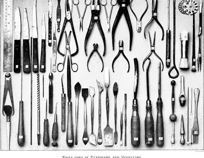
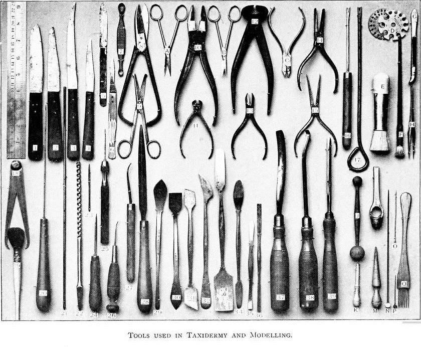
Taxidermist’s tools.  A taxidermist’s tools may seem nightmarish, but each served a valuable purpose, such as Tool No. 1, a skinning knife, and Tool No. 7, the “the eye and brain scoop or extractor—a tool which may save the beginner some trouble in preventing the bursting of a bird’s eyeballs” (20).

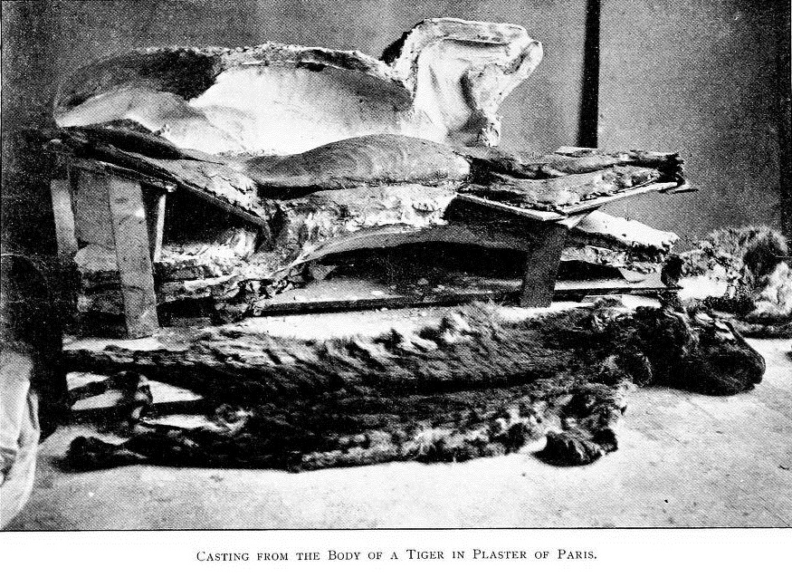
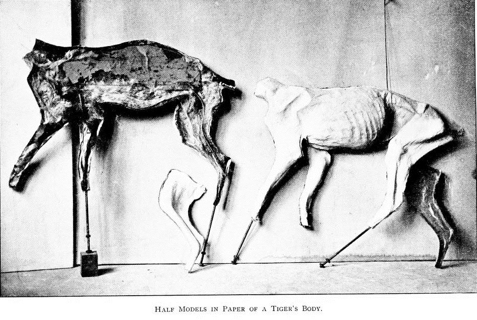
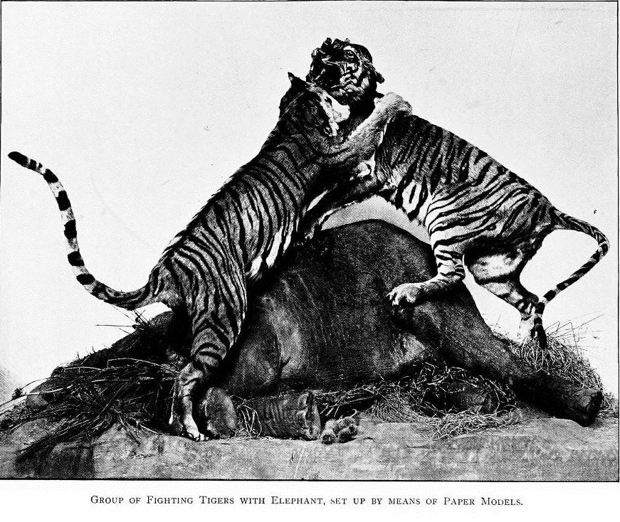
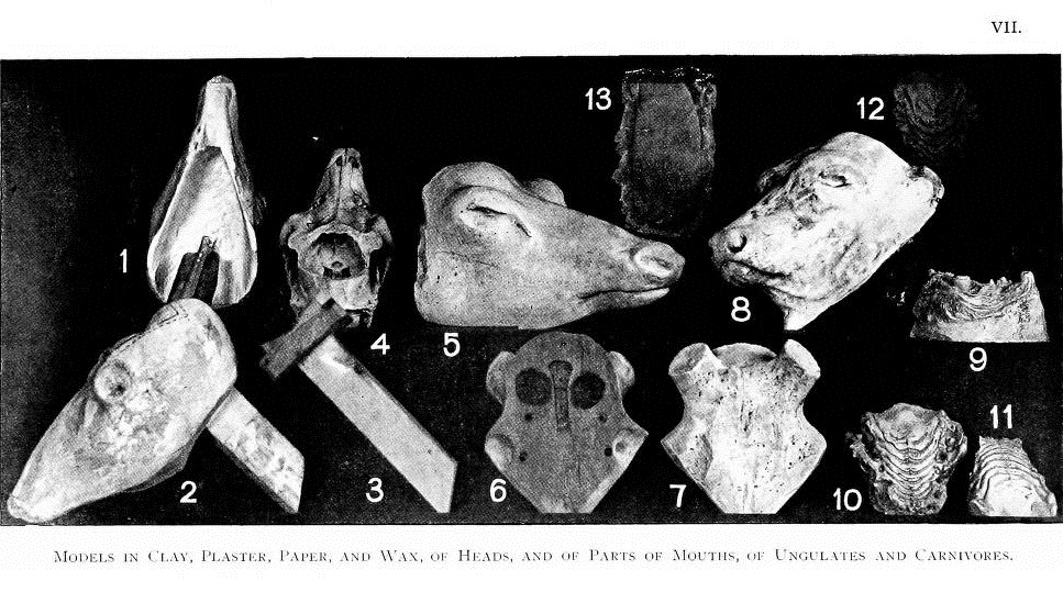
Despite the less pleasant aspects involved in taxidermy, it does require practitioners with real artistic talent capable of accurately sculpting a model of whichever vertebrate animal has been skinned (invertebrates require other methods of preservation).  Browne describes the process of taking plaster casts of the animal, useful for modeling small details like the palatal ridges inside the mouth of a tiger (123).  Casts of skinned animals also allow the taxidermist to create accurate under-molds upon which to arrange the flayed and preserved skin; the taxidermist will press torn pieces of paper covered in glue into the hollow, two-piece mold in layers, in order to create the under-form.  Browne describes this process:

“A half-bucketful of flour-paste was made, and some thin but tough waste-paper, used by manufacturers in the hosiery trade, was procured.  This was torn into pieces of various sizes, and taking one—pasted on one side only—sufficient for the purpose, a commencement was made at one end by laying the unpasted side downwards, and working it into the inequalities of the mould by gentle but even pressure with the fingers, the edge of the paper coming over the edge of the mould being pasted down on the flat in order to get a secure attachment.  Piece by piece the whole of the inside was covered” (132).

After the two halves of the plaster mold were filled with glued paper, the mold hardened, and the halves were fastened together.  The animal’s skin was draped over the mold and parts like the feet were filled with clay, the tail with a rod (137), and visible muscles of the jaw with colored wax (138).  Parts of the skin (such as the exterior of padded feet) were waxed to appear natural, and artificial eyes “made of half-ovals of thin glass painted to nature from the inside” were “inserted in the orbits after the eyelids have been filled, and the cornea, etc., then made up in wax” (139).

View Full Size Image
Casting the skinned body of a tiger.

View Full Size Image
Paper under-mold of a tiger, in two halves.

View Full Size Image
Taxidermy of tigers fighting over an elephant, done in the manner described by Browne above.

View Full Size Image
Models of animal heads, made in various different media, used to wrap the animals’ skins around.

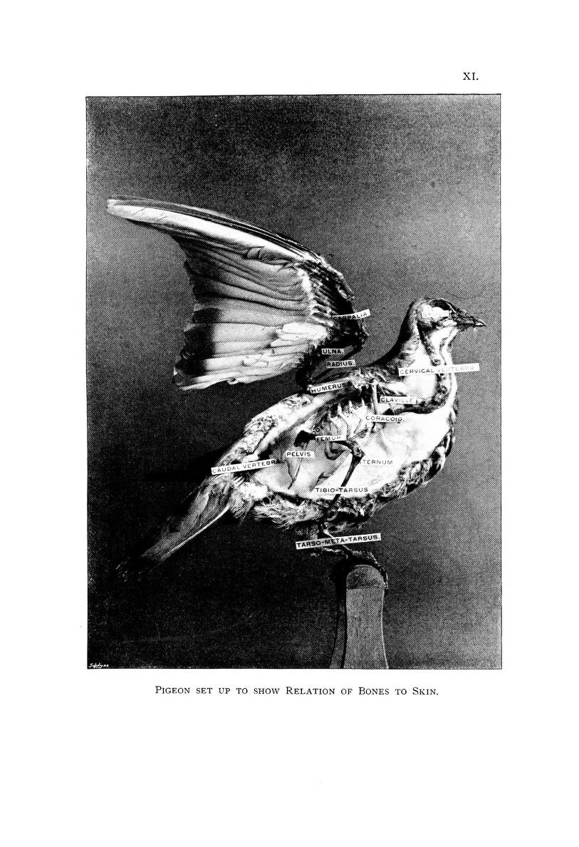
While their methods may seem gruesome to some, taxidermists have created lasting models that represent not only lifelike specimens, but also teaching specimens that carefully show the physiological structure of different animals.  In the illustration below, a pigeon has been preserved to show the relation of its bones to its skin.

View Full Size Image
Taxidermy of a pigeon showing the relation of its bones to skin.

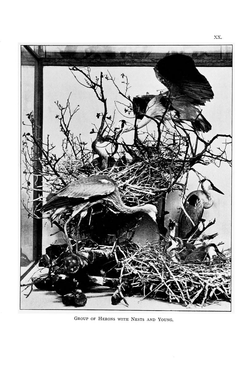
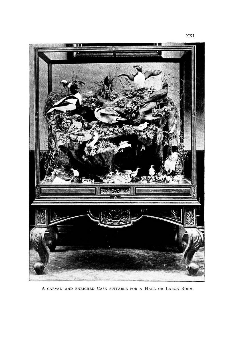
Also part of the taxidermist’s artistry is his or her ability to display the specimens in settings that look like their natural habitat, and to do so in an aesthetically pleasing manner.  Browne treats this subject, and the photographs below are examples of beautifully arranged preserved specimens.

View Full Size Image
Taxidermy of several Herons.

View Full Size Image
Taxidermy of a group of penguins.

Whatever your opinions of taxidermy, it has been crucial to building the collections of many museums we know and love, and to helping our understanding of many different animal species.

July 3, 2013by jjones
Blog Reel, Featured Books

Death by Corset: A Nineteenth-Century Book about Fatal Women’s Fashions (and Animal Physiology)

Normally at the BHL we examine zoology-related books in our Book of the Week posts, but this week we’re entering new territory and taking a look at a comparative physiology book that examines the only species to mutilate its own body: Homo sapiens.

If you go by what J. L. Comstock, M.D., had to say in 1848 about what it was like to be a lady, times were difficult.  In this week’s book of the week, Outlines of Physiology, both Comparative and Human; in which are Described the Mechanical, Animal, Vital, and Sensorial Organs and Functions; Also, The Application of These Principles to Muscular Exercise, and Female Fashions and Deformities, Comstock explains that young women were under enormous pressure to be aesthetically pleasing to men (not a bad goal, he points out), and this pressure to look good by wearing a corset or stays was causing rampant deformities, illness, and even death: “. . .I have no doubt that the ladies themselves, to a considerable extent, will agree with me in believing, that hundreds, nay thousands, of females literally kill themselves every year by this fashion in our own country: and if suicide is a crime, how will such escape in the day of final account!” (311).

While he may have thought that women who died of tight-laced corsets were going to hell, he did sympathize with their goal to look beautiful for men: “what possible motive could have induced the females of the present age, and especially those of these United States (where ultraism in respect to this deformity [by corset] is carried to a much greater extent than in any other country). . .Can it be for the purpose of making themselves more agreeable, and more acceptable to the lords of creation?  Then certainly their motives ought to meet with the law of kindness, and the tortures through which they are willing to pass in order to arrive at perfection—the sympathy and commiseration of those for whom such perils are encountered” (321).
View Full Size Image

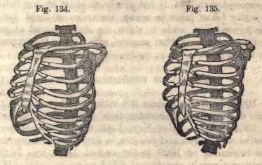
Comstock explains: “We have represented by figures 134 [normal skeleton] and 135 [deformed skeleton] the difference between a natural human skeleton, and one in which the pressure of stays has pushed the front ends of the ribs inward, bending the soft cartilages, so as to make them form acute angles outward” (311-312).  This results in the ribs pressing inward upon the lungs.
View Full Size Image

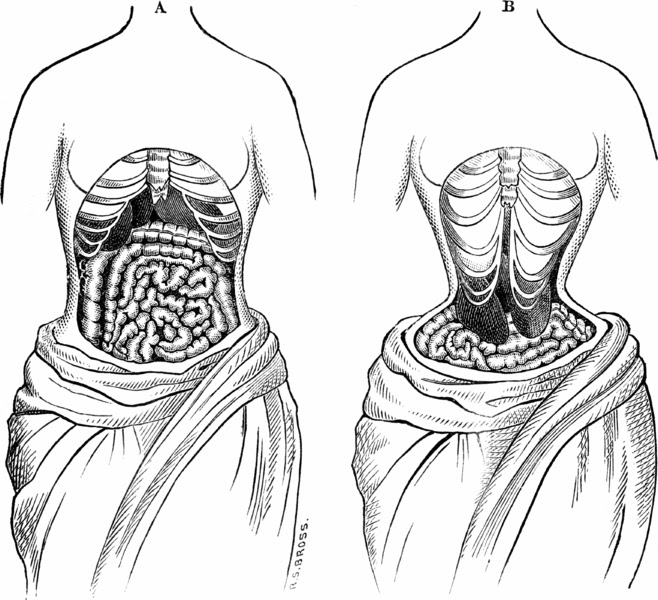
The force of a tight-laced corset upon the ribs caused them to create pressure not only upon the lungs, but also the other internal organs which undergo a shifting in position to accommodate for the new skeletal shape.  This illustration from 1884 represents the contemporary medical opinion about organ displacement due to corset wearing.

Ann Buermann Wass and Michelle Webb Fandrich, two fashion historians, describe what corsets looked like during Comstock’s period:

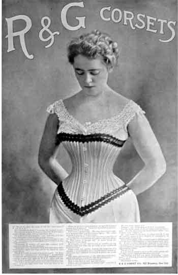
“By the 1840s, the line of women’s corsets extended to well below the waist. The corset of the 1840s and 1850s was boned throughout with whalebone and, as it had been in decades prior, outfitted with a metal, wood or whalebone busk at the center front. Commenting on the gradual lengthening of the corset in 1841, the Handbook of the Toilet noted that “[t]he modern stay extends not only over the bosom but also all over the abdomen and back down to the hips…they have been growing in length by degrees”. . .By this time, corsets had lost their shoulder straps and were most commonly fastened up the front with busk outfitted with clips and laced up the back to achieve a more precise fit.” (306)
View Full Size Image

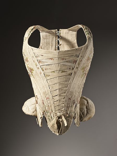
An early example of a stay, a type of corset with shoulder straps made of thick material.  This particular example is a woman’s corset from France, c. 1730-1740, made in a silk plain weave with supplementary weft-float patterning.  From the Los Angeles County Museum of Art.
View Full Size Image

A corset ad from 1898 developed by Earnest Elmo Calkins for R & G Corset Company.  This corset represents the longer shape described by Wass and Fandrich.

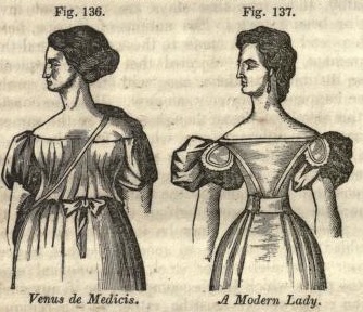
The deformities caused by stays and corsets were a serious problem for Comstock, not only because they could result in death but because he believed that children born of corset-wearers were weak, small and sickly; this is owing to Comstock’s belief that corsets deplete the mother’s milk supply: “[the breasts of a corset- or stay-wearer] present a mere pretence, a nullity, a source of starvation, rather than one of sustenance, to the nascent beings, who are so unfortunate as to be thrown upon such cotton and wool resources of existence” (320-321).
View Full Size Image

Figure 136 represents an ideal woman who does not wear a corset or stays and can thus produce breast milk and strong children.  Figure 137 represents the typical woman contemporary to Comstock who wears stays and thus cannot produce breast milk or healthy children, according to the M.D. (320).

And shoulder-baring dresses?  Ladies, forget about it!  Comstock notes that:

“the] cause of distorted shoulders. . .is the recent fashion of dressing so wide across the neck as to leave one, or perhaps both the acromion processes, or shoulder tips, in a state of entire nudity. . .The consequence of this fashion was, that, judging from the perpetual motion of [the shoulders], the wearer constantly felt as though her dress was in danger of slipping down, and which she made as constant efforts to prevent, or to ascertain by feeling with the shoulder whether this was the case, or not, until these motions became habitual, and therefore insensible. . .until [one shoulder] became permanently higher than the latter.” (296-7)

Comstock’s only hope to ending the serious deformities and risk for death caused by stays, corsets and fashionable clothing was that the country simpletons who didn’t know that corsets were fashionable would move to the city and literally create a new race of revitalized humans: “It is true that there are parts of our country where the practice of excessive lacing, and therefore its degenerating consequences do not exist; and whence we are happy to know that many of the daughters of unsophisticated nature are transplanted into our cities, there to become the fostering angels of a renovated species” (321).

While Comstock also explores similarities between human and animal physiologies in his book, a large part of his focus is on remedies for men and women to avoid illness.  Among Comstock’s recommendations for different forms of “exciting exercise” for adults and children to prevent illness are “field-sports,” angling, riding, calisthenics and archery—but also “cheerfulness” and “laughing” (239-266).
View Full Size Image

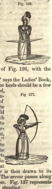
And ladies, while he doesn’t want you to wear a corset during your archery practice, don’t forget to wear your long-sleeve, floor-length gown with giant feathered hat (270).

If you are sitting at a desk right now while you read this, beware that you don’t become the lazy, deformed, lolling, school-girl type described by Comstock in his section on the evils of the seated posture:

“One posture which school-girls are exceedingly apt to take, is that of leaning forward, and placing the elbow on the desk for support; and this they often do, even when their seats are provided with backs.  This posture, if continued so as to form a habit, will often show its effects on all occasions, the young lady having such a disposition to lean, as to indulge it when any support happens to be near where she sits, let the place or company be what it may.  Such a one will lean, with the hand supporting the head, when at home, on a table, or window-stool, or any other convenient lolling-place, for hours together.” (294)
View Full Size Image

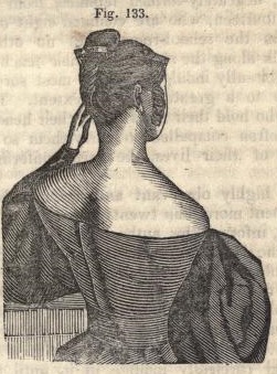
If you spend your time leaning with your head in your hand while sitting, you can look forward to deformed shoulder blades, according to Comstock, “one of which is sometimes thrown so far out of place as to give it the appearance of absolute dislocation” as in Figure 133 (294-295).

While Comstock’s pronouncements and prescriptions may seem amusing, corsets and stays presented a very real danger in the minds of many of his contemporaries.  Comstock’s descriptions of the physiological changes caused by corsets were sometimes accurate, such as his description of compressed lungs and organs, which resulted in difficulty breathing and could make recovery from any underlying illness harder.  Valerie Steele, a fashion historian, explains 19th-century beliefs about the dangers of corsets:

“The corset has been blamed for causing dozens of diseases, from cancer to curvature of the spine, deformities of the ribs and displacements of the internal organs, respiratory and circulatory diseases, birth defects, miscarriages, and “female complaints,” as well as medical traumas such as broken ribs and puncture wounds.  The Lancet, Britain’s most important medical journal, published more than an article a year from the late 1860s to the early 1890s on the medical dangers of tight-lacing.  Deaths from tight-lacing were also mentioned.  In one case, for example, the “heart was found to be so impeded in its action as to render life impracticable.” (67)

Today, corsets are still in fashion among people interested in recreating historical costume, and also among various different types of lingerie enthusiasts.  Regardless of one’s beliefs about the safety or practicality of corsets, they present a fascinating reflection of human beliefs about beauty and the ability to manipulate the human body.

References:

Comstock, J.L. Outlines of Physiology, both Comparative and Human; in which are Described the Mechanical, Animal, Vital, and Sensorial Organs and Functions; Also, The Application of These Principles to Muscular Exercise, and Female Fashions and Deformities.  New York: Pratt, Woodford & Company, 1898.

Steele, Valerie.  The Corset: A Cultural History.  New Haven, CT: Yale University Press, 2003.  p. 67.

Wass, Ann Buermann and Michelle Webb Fandrich.  Clothing through American History: The Federal Era through Antebellum, 1786-1860.  Santa Barbara, CA: Greenwood, 2010.  p. 306.

June 27, 2013by jjones

Help Support BHL

BHL's existence depends on the financial support of its patrons. Help us keep this free resource alive!

search

About BHL

The Biodiversity Heritage Library (BHL) is the world’s largest open access digital library for biodiversity literature and archives. BHL operates as a worldwide consortium of natural history, botanical, research, and national libraries working together to digitize the natural history literature held in their collections and make it freely available for open access as part of a global “biodiversity community.”

Join Our Mailing List

Sign up to receive the latest news, content highlights, and promotions.

Subscribe Now

Subscribe to Blog via Email

Enter your email address to subscribe to this blog and receive notifications of new posts by email.

Join 319 other subscribers

Subscribe to Blog Via RSS

Subscribe to the blog RSS feed to stay up-to-date on all the latest BHL posts.

Access RSS Feed

Inspiring Discovery through Free Access to Biodiversity Knowledge.

The Biodiversity Heritage Library makes it easier than ever for you to access the information you need to study and explore life on Earth…for free, anytime, anywhere.


64+ Million Pages of
Biodiversity Literature Online.

EXPLORE

Tools and Services
to Transform Research.

EXPLORE

300,000+
Illustrations on Flickr.

EXPLORE

ABOUT | HARMFUL CONTENT | PRIVACY | SITE MAP | TERMS OF USE