Biodiversity Heritage Library - Program news and collection highlights from BHL
  • Home
  • News
  • Featured Books
    • All Featured Books
    • Book of the Month Series
    • BHL at 20
  • User Stories
  • Campaigns
    • Fossil Stories
    • Garden Stories
    • Monsters Are Real
    • Page Frights
    • Her Natural History
    • Earth Optimism 2020
  • Tech Blog
  • Visit BHL
Home
News
Featured Books
    All Featured Books
    Book of the Month Series
    BHL at 20
User Stories
Campaigns
    Fossil Stories
    Garden Stories
    Monsters Are Real
    Page Frights
    Her Natural History
    Earth Optimism 2020
Tech Blog
Visit BHL
  • Home
  • News
  • Featured Books
    • All Featured Books
    • Book of the Month Series
    • BHL at 20
  • User Stories
  • Campaigns
    • Fossil Stories
    • Garden Stories
    • Monsters Are Real
    • Page Frights
    • Her Natural History
    • Earth Optimism 2020
  • Tech Blog
  • Visit BHL
Biodiversity Heritage Library - Program news and collection highlights from BHL
Blog Reel, User Stories

Tasting Platypus Milk: Linking Specimens and Stories

illustrations of the platypus

Zoological knowledge typically comes from one of two primary sources: the living and the dead — observations of animals going about their business in their habitats; and the study of preserved specimens. We rarely get the whole picture of an animal’s natural history without both, and each feed into how species are portrayed to those that have never seen them.

Getting echidnas wrong

A1.2 7_Tachyglossus_aculeatus.JPG

Taxidermy short-beaked echidna (Tachyglossus aculeatus) from the University Museum of Zoology, Cambridge. Note the backwards-pointing hind feet. Specimen A1. 2/7. Photo Credit: University of Cambridge

Consider the echidna, for example. Examining echidna specimens in museums helps us to understand their evolutionary relationships and enables us to come up with some reasonable suggestions for how they live their lives. The impenetrable coat of thick spines is clearly for defense against predators and guessing at an ant-eating lifestyle doesn’t require too much imagination when you think about what its narrow toothless snout and stout, digging claws might be for. However, we wouldn’t be able to confirm exactly how these things are put to use without seeing them alive.

As someone with one foot in the world of Australian mammal ecology (the living) and another in natural history museums (the dead), one thing that really interests me is the cross-over: how do the dead represent the living? Museums are sites of communication — they provide windows onto the natural world for people to engage with animals they may never see alive. This has made the topic of how animals are depicted — in writings, illustrations and museums — a real focus for me.
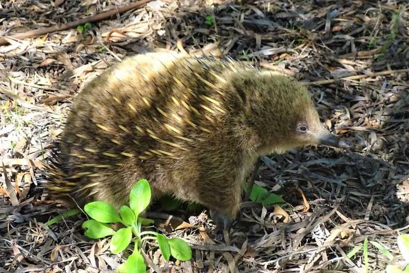
Bruny Island 8 (16) Ashby.JPG

A live (undoctored) echidna, in Tasmania. Photo Credit: Jack Ashby.

While museum specimens are in one sense a primary source, they are also modified by people in order to make them presentable to the public. Taxidermy is intended to make visitors forget the animal is dead, and to achieve that a flat animal skin has to be rebuilt into the shape of a living animal. The trouble is that throughout history, the person doing the taxidermy — particularly for specimens that had travelled to Europe from Australia — had often never seen the living animal. This means that their poses are often incorrect.

In the case of the echidna, it is extremely common for the back feet to point in the wrong direction — in life they should point backwards (enabling echidnas to scratch in between their spines and dig vertically downwards to bury themselves in defense), but taxidermists often didn’t know that, so twisted their feet around. I’ve come across tens of wrong-footed echidnas, and I know the reason why, but I was flummoxed a few years ago when I encountered an echidna with blue eyes at the Manchester Museum. Where did that error come from?
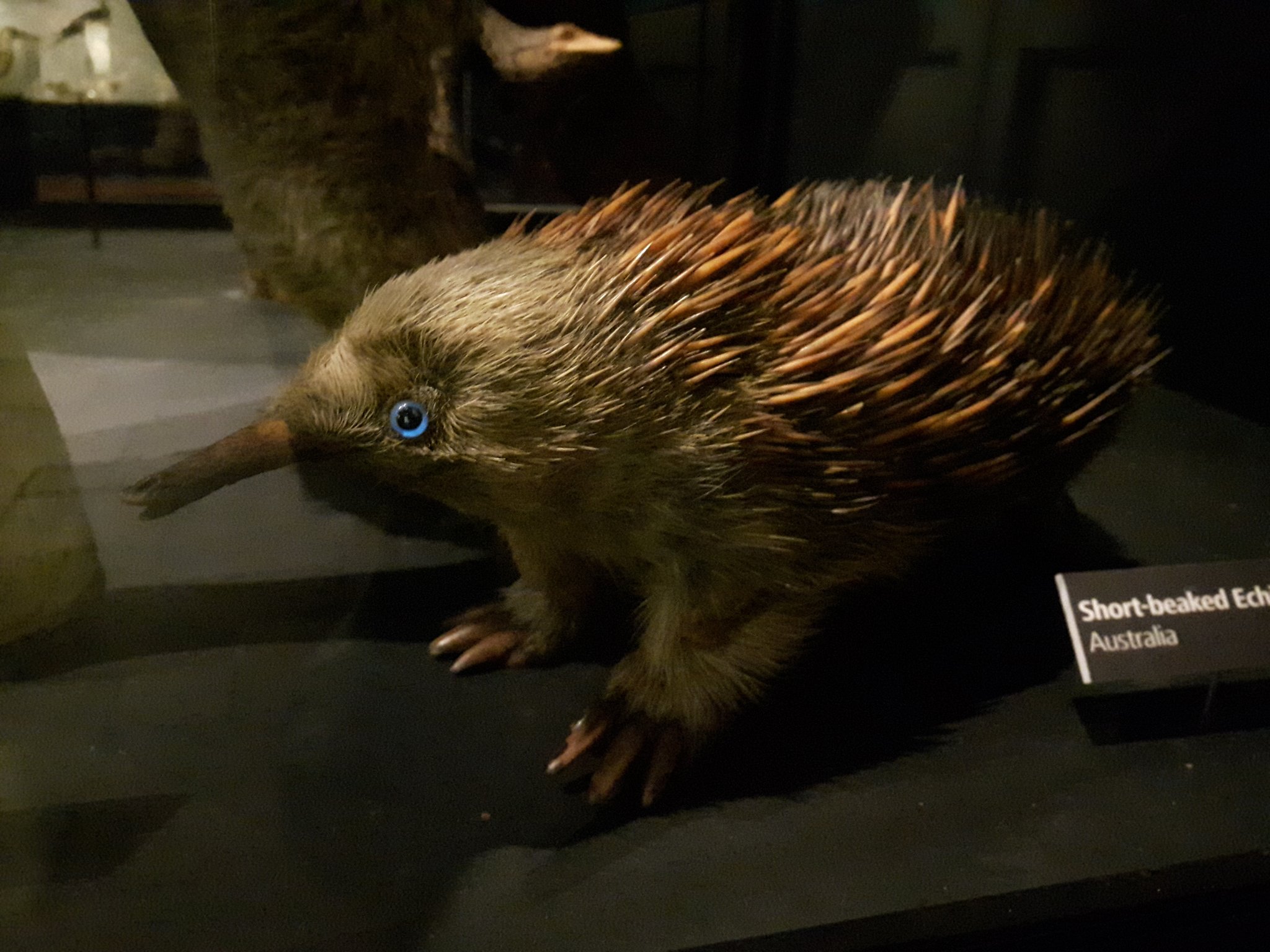
Manchester.jpg

Why does this taxidermy echidna at Manchester Museum have blue eyes? Photo Credit: Jack Ashby.

That mystery echidna popped back into my mind recently when re-reading the original species description on the Biodiversity Heritage Library, and I think I know the answer.

In 1792 George Shaw (who would also publish the first scientific description of the platypus seven year later) included “The Porcupine Ant-Eater” in The Naturalist’s Miscellany. Amongst an otherwise perfectly reasonable description of the species (which he considered to provide a link between actual porcupines in the genus Hystrix and actual ant-eaters in the genus Myrmecophaga), Shaw includes this line:

“The snout is long and tubular, and perfectly resembles in structure that of the Myrmecophaga jubata, or great ant-eater; having only a very small opening or rictus at the tip, from whence is protruded a long lumbriciform [worm-like] tongue, as in the ant-eaters. The nostrils are small, and seated near the extremity of the snout. The eyes are very small, and black, with a pale-blue iris.”
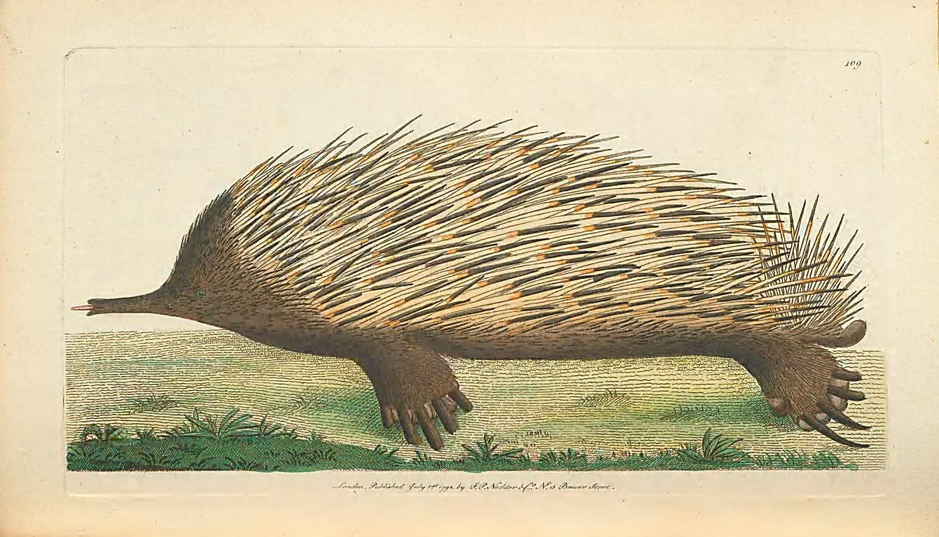
naturalistsmisc3shaw_0253.jpg

Plate 109. Shaw, George. The naturalist’s miscellany. Volume iii. 1792. Contributed in BHL by Museums Victoria.

Curiously, the illustration that accompanies the text (above) — which is assumed to be based on a painting by Sydney-based collector John White — has correctly coloured black eyes. The question then becomes why Shaw thought echidnas had blue eyes — could it be that the eyes of Shaw’s specimen (sent by Joseph Banks) had been discoloured by the preservative? In any case, it seems likely that the echidna in Manchester was modified to fit Shaw’s original description.

Elsewhere on BHL, we can find examples of illustrations of echidnas that were clearly based on doctored dead specimens, rather than observations of living ones, as their feet incorrectly point forwards and the animals appear flattened. Artists were basing their depictions on “primary sources” in museum collections, but unfortunately those specimens weren’t accurate.
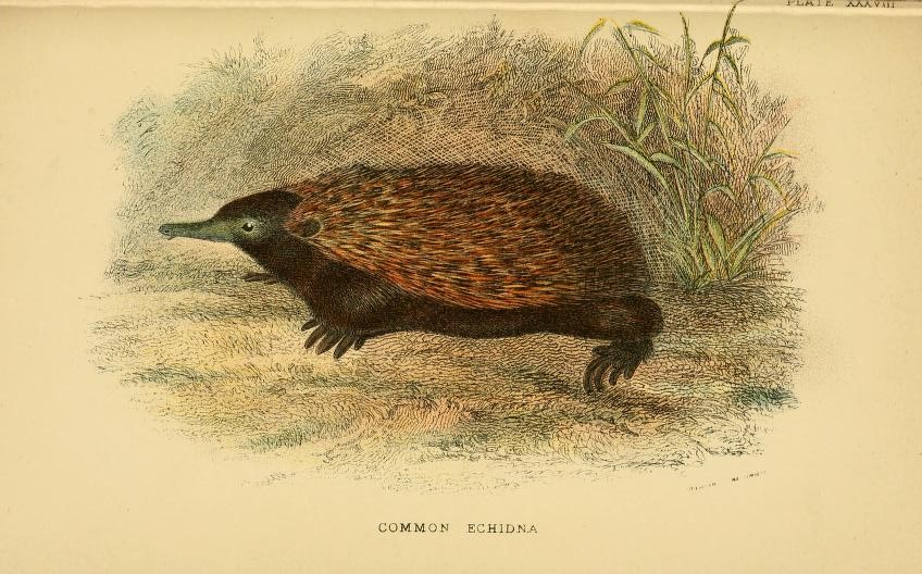
handbooktomarsup00lydekk_0339.jpg

Plate XXXVIII, an echidna with forward-facing feet. Lydekker, Richard, A hand-book to the marsupialia and monotremata. 1896. Contributed in BHL from Smithsonian Libraries.

Tasting platypus milk

I am currently writing a new book on the echidnas’ relative, the platypus. I’ve been utterly reliant on the incredible collection of works on BHL to tell the story of the importance of platypus representations on the history of taxonomy. The introduction of the species to scientific circles in Europe sparked a decades-long debate over where it fitted on the tree of life and what characteristics mammals could possess. One of the many arguments in contention was whether or not they are mammals, another was whether or not they lays eggs. We now know they are, and they do.

A defining feature of mammals is that they feed their young with milk, so those who argued that platypuses are mammals were searching for evidence of milk-producing mammary glands even though platypuses lack nipples. Richard Owen was among those who was attempting to prove their milkiness.

The battle is played out in the documents available on BHL, and to my absolute delight, it led me to this line in one of Owen’s papers on platypus anatomy. Owen had squeezed what he thought was the mammary gland of a preserved platypus and watched the holes where the glands opened onto the skin:

“[T]here escaped from these orifices minute drops of a yellowish oil, which afforded neither perceptible taste nor smell, except such as was derived from the preserving liquor”.

Owen was so dedicated to his argument that he tasted the milk from a platypus that had been dead for many months, or even years.

Without a resource like BHL, putting together the pieces of the platypus puzzle across 220 years, and the changing way that specimens and field-observations have been used to inform representations of the animal over time, would be extremely difficult.
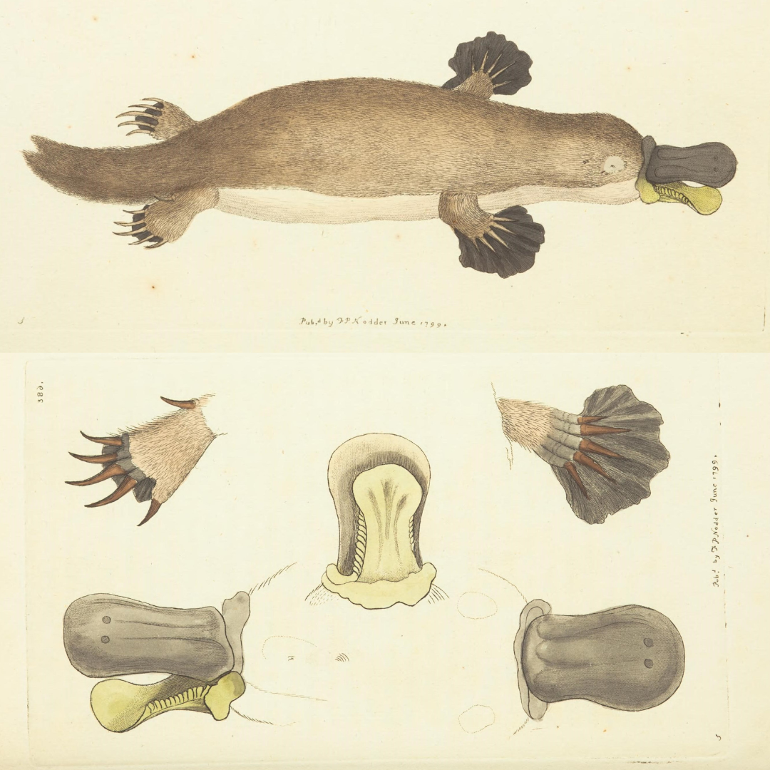
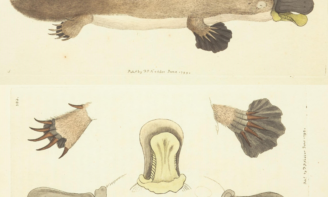
illustrations of the platypus

George Shaw, who provided the first scientific description and published illustration of the platypus (Ornithorhynchus anatinus) in 1799 within The Naturalist’s Miscellany, wrote: “… it naturally excites the idea of some deceptive preparation by artificial means …”. Shaw, George. The naturalist’s miscellany. v. 10 (1799). Contributed in BHL from Museums Victoria. DOI: https://doi.org/10.5962/p.304567.
July 7, 2020by Joel Richard
Blog Reel, Featured Books

To Contemplate Without Dread: Nineteenth Century Taxidermy and the Study of Natural History

Natural history illustrations often aim to show life-like flora and fauna. Depictions of birds poised to take flight, fish swimming upstream, and mammals mid-stride are common in 18th and 19th century zoology and botany publications. What is lost in these often lavish illustrations is a certain truth about the way many naturalists interacted with their objects of study: many species of plant or animal were first encountered not in the wild, but in the display case, in the form of carefully prepared specimens.

One British naturalist, Captain Thomas Brown (1785-1862), made the practical observation that mounted animal specimens allowed naturalists to “contemplate, without dread, the most destructive and furious quadrupeds, and the most noxious reptiles.” It follows that many dangerous and rare species may not have been studied if not for taxidermy. Similarly, the building up of large and comprehensive natural history collections would not have been possible without a means of preserving specimens for the long term. As early collectors and naturalists quickly realized, proper taxidermy techniques were necessary to ensure that museum and cabinet collections did not degrade and disintegrate over time. Thus, a significant part of natural history is the rather unnatural history of taxidermy and specimen preparation.

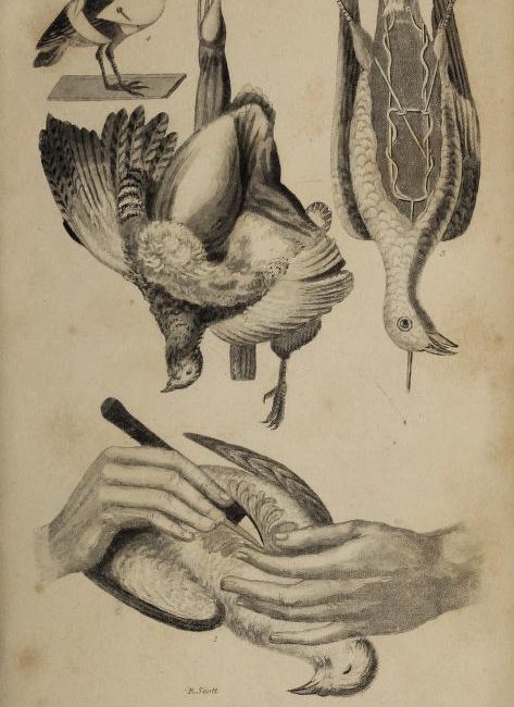
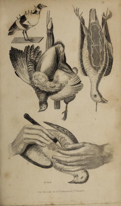
taxidermistsmanu00brow_0_0021.jpg

Plate III. Brown, Thomas. The Taxidermist’s Manual, or, The Art of Collecting, Preparing and Preserving Objects of Natural History. 1833.  Contributed in BHL by The Thomas Fisher Rare Book Library, University of Toronto.

While elements of modern taxidermy can be traced back to ancient Egyptian embalming practices, taxidermy as a tool for the study of natural history did not come about until the mid-18th century. French entomologist René Antoine Ferchault de Réaumur’s treatise, ‘Divers means for preserving from corruption dead birds intended to be sent to remote countries, so that they may arrive there in good condition…‘, published in the Transactions of the Royal Society in 1748, is often cited as the first comprehensive discussion of the topic. However, Réaumur proposed techniques for preparing specimens for shipment only, not long-term preservation or display.

More comprehensive techniques developed in the 19th century into what many referred to as the ‘Art’ of taxidermy, and it was during this time that popular printed manuals on taxidermy proliferated. The Biodiversity Heritage Library currently includes over fifty examples of such manuals, published between 1799 and 1927.

Captain Thomas Brown, better known for his work in the field of conchology (the species of sea-snail Zebina browniania is named after him), published The Taxidermist’s Manual, or, The Art of Collecting, Preparing and Preserving Objects of Natural History in 1833. The work covers in rather gruesome detail the methods for skinning and preparing a range of mammals, fish and birds – including species as large as the elephant – as well as techniques for mounting smaller crustacea and insects. It includes “Mr. Waterton’s method for stuffing birds,” referring to the more famous naturalist Charles Waterton (1782-186), gives recipes for chemical compounds used in preservation, describes two methods for constructing false eyes out of enamel, and lists common prices for some of the supplies required. The manual also gives creative advice for mounting and positioning each species: sloths, for example, should always be mounted in an upright position, as they are seldom seen walking horizontally.

Brown’s general advice to the taxidermist is “copy nature whenever you have it in your power.”

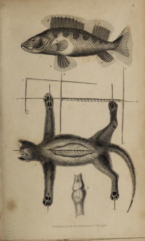
taxidermistsmanu00brow_0_0019.jpg

Plate II. Brown, Thomas. The Taxidermist’s Manual, or, The Art of Collecting, Preparing and Preserving Objects of Natural History. 1833. Contributed in BHL by The Thomas Fisher Rare Book Library, University of Toronto.

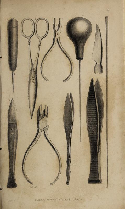
The text is illustrated by plates cut by Scottish engraver Robert Scott. In stark contrast to the life-like creatures often found in many illustrated natural history volumes, the animals here (including what appears to be a poor domestic cat) are very clearly dead, though they are still idealized. More macabre images may be found on plates four and five, which show the various surgical instruments – scalpels, mallets and pliers – used in the various stages of animal taxidermy. Perhaps the strangest inclusion, however, is a short appendix in which Brown describes a newly discovered French technique for preserving the human body.

taxidermistsmanu00brow_0_0025.jpg

Plate VI. Brown, Thomas. The Taxidermist’s Manual, or, The Art of Collecting, Preparing and Preserving Objects of Natural History. 1833.  Digitized by The Thomas Fisher Rare Book Library, University of Toronto.

Popular works like Thomas Brown’s Taxidermist’s manual reveal in morbid detail the unnatural materials and practices that coincided with the study of natural history – the arsenic, wire, stuffing, and false eyes that naturalists employed in their attempts to “avert the process of Time’s destroying hand.”

October 25, 2018by Joel Richard
Blog Reel, Featured Books

Taxidermy: The Artistry of Preserving Bodies

Thanks to reality television shows like “Oddities” on the Science Channel, “Immortalized” on AMC, and “American Stuffers” on Animal Planet, there has been a resurgence in the popularity of taxidermy.  The first instance of a dead organism being preserved and stuffed for display or learning purposes is not known with certainty, but in our book of the week, we’ll learn several theories on the origins of taxidermy—and most interestingly, how to perform it.  Montagu Browne discusses all aspects of taxidermy, and its origins, in his 1896 work, Artistic and Scientific Taxidermy and Modelling: A Manual of Instruction in the Methods of Preserving and Reproducing the Correct Form of All Natural Objects, Including a Chapter on the Modelling of Foliage:

“Little is known of the beginnings of the practice of the ‘stuffing’ or ‘setting-up’ of animals for ornament or for scientific purposes; and it is highly probable, from what we gather from old works of travel or natural history, that the art is not more than some three hundred years old.  It was practiced in England towards the end of the seventeeth century, as is proved by the Sloane collection, which in 1725 formed the nucleus of the collection of natural history now lodged in the galleries at South Kensington.

…the oldest museum specimen in existence is a rhinoceros still preserved in the Royal Museum of Vertebrates in Florence.  This was for a long time a feature of the Medicean Museum in Florence, and was originaly mounted for the museum of Ulysses Aldrovandus in Bologna.  It dates from the sixteenth century.” (3

Browne says that the first taxidermy performed upon birds took place when, in the early sixteenth century, Holanders began commercial trade with the East Indies.  He says:

“A nobleman brought back to Amsterdam a large collection of live tropical birds and placed them in an aviary, which was heated to the proper temperature by a furnace.  It happened that the attendant one night before retiring carelessly left the door of the furnace open, thereby allowing the smoke to escape, which suffocated the birds.  The nobleman beholding the destruction of his large collection, which was the pride of the city, began to devise means for the preservation of the dead birds.  To this end the best chemists of Amsterdam were called in for consultation, and it was decided to skin the birds and fill their skins with the spices of the Indies for their preservation.  This was done, and they were then wired and mounted to represent life.” (3)

While taxidermy results in the creation of beautiful, preserved biological specimens, there is the gruesome aspect of needing to capture and kill these organisms prior to preservation (if they have not died naturally).  Browne dedicates many pages to the various means of killing different organisms, both vertebrate and invertebrate, so that their bodies remain intact and suitable for taxidermy.  These methods are not pretty, from soaking invertebrates in different mixtures of sea water, alcohol, acids and nicotine, to using cyanide of potassium and chloroform on vertebrates (60), or setting traps for them or hunting them (95).

The business of flaying an animal and preserving its skin can be complex depending on the animal’s size, and Browne quotes M. Dufresne’s account of skinning and preserving an elephant that had died at the menagerie of the Jardin des Plantes in Paris in 1803:

“The corpse of the elephant having been extended upon the ground, facilitated our taking and writing all its dimensions. . .M. Desmoulins drew the animal on one of the sides of the wall according to all these measurements, in the workshop where the model was to be constructed in its natural size.  This done, we proceeded to the skinning of the elephant, which we were only able to place upon its back by means of four-corded pulleys, fastened to the platform.  In this position we made an incision in the form of a double cross: the middle line went from the mouth to the anus, the two others were directed from each left foot to the opposite right foot; the tail and trunk were opened underneath longitudinally.  We scooped out the soles of the feet within an inch of their edge, that the nails might remain in the skin: to effect this, we were obliged to employ the chisel and mallet.  This operation was very difficult.

After four days’ labour of several persons, we separated the skin from the body: it then weighed 576 lbs; we extended it on the ground to take away the cutaneous muscles which adhered to its interior, particularly to the head” (117).

Dufresne goes on to describe putting the skins in tubs of chemicals and boiling water, and then placing the skins over a life-size model of an elephant made of hollow wooden pieces (118).  After many days of work, the entire process was completed, and the elephant skin was rubbed with turpentine and olive oil to restore its original color after it had been made “a very ugly grey colour” from the chemicals in which it had soaked (119).

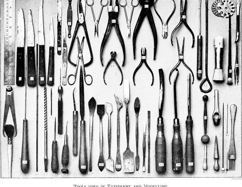
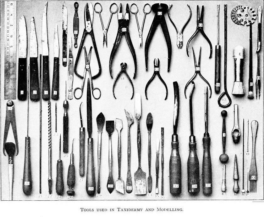
View Full Size Image

Taxidermist’s tools.  A taxidermist’s tools may seem nightmarish, but each served a valuable purpose, such as Tool No. 1, a skinning knife, and Tool No. 7, the “the eye and brain scoop or extractor—a tool which may save the beginner some trouble in preventing the bursting of a bird’s eyeballs” (20).

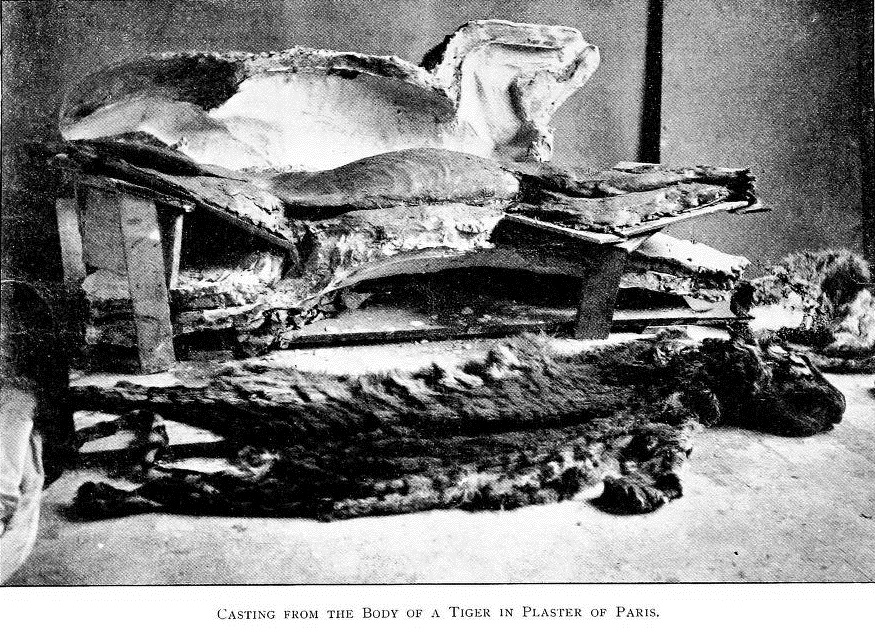
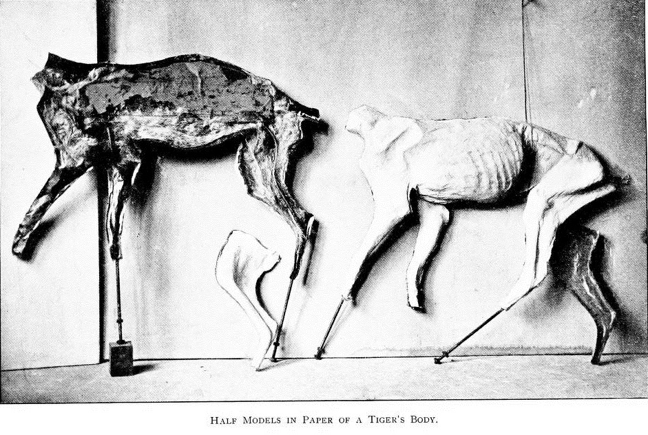
Despite the less pleasant aspects involved in taxidermy, it does require practitioners with real artistic talent capable of accurately sculpting a model of whichever vertebrate animal has been skinned (invertebrates require other methods of preservation).  Browne describes the process of taking plaster casts of the animal, useful for modeling small details like the palatal ridges inside the mouth of a tiger (123).  Casts of skinned animals also allow the taxidermist to create accurate under-molds upon which to arrange the flayed and preserved skin; the taxidermist will press torn pieces of paper covered in glue into the hollow, two-piece mold in layers, in order to create the under-form.  Browne describes this process:

“A half-bucketful of flour-paste was made, and some thin but tough waste-paper, used by manufacturers in the hosiery trade, was procured.  This was torn into pieces of various sizes, and taking one—pasted on one side only—sufficient for the purpose, a commencement was made at one end by laying the unpasted side downwards, and working it into the inequalities of the mould by gentle but even pressure with the fingers, the edge of the paper coming over the edge of the mould being pasted down on the flat in order to get a secure attachment.  Piece by piece the whole of the inside was covered” (132).

After the two halves of the plaster mold were filled with glued paper, the mold hardened, and the halves were fastened together.  The animal’s skin was draped over the mold and parts like the feet were filled with clay, the tail with a rod (137), and visible muscles of the jaw with colored wax (138).  Parts of the skin (such as the exterior of padded feet) were waxed to appear natural, and artificial eyes “made of half-ovals of thin glass painted to nature from the inside” were “inserted in the orbits after the eyelids have been filled, and the cornea, etc., then made up in wax” (139).

View Full Size Image
Casting the skinned body of a tiger.

View Full Size Image
Paper under-mold of a tiger, in two halves.

View Full Size Image
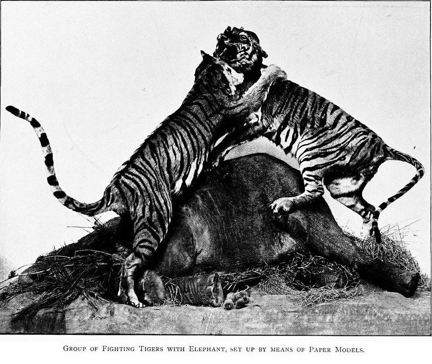
Taxidermy of tigers fighting over an elephant, done in the manner described by Browne above.

View Full Size Image
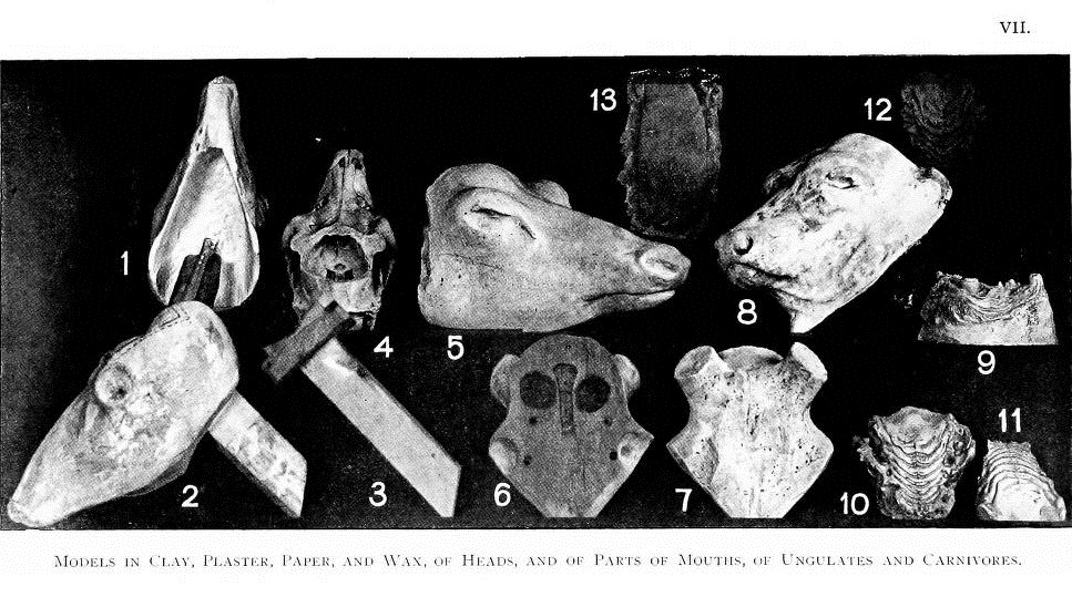
Models of animal heads, made in various different media, used to wrap the animals’ skins around.

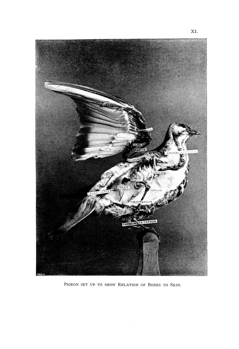
While their methods may seem gruesome to some, taxidermists have created lasting models that represent not only lifelike specimens, but also teaching specimens that carefully show the physiological structure of different animals.  In the illustration below, a pigeon has been preserved to show the relation of its bones to its skin.

View Full Size Image
Taxidermy of a pigeon showing the relation of its bones to skin.

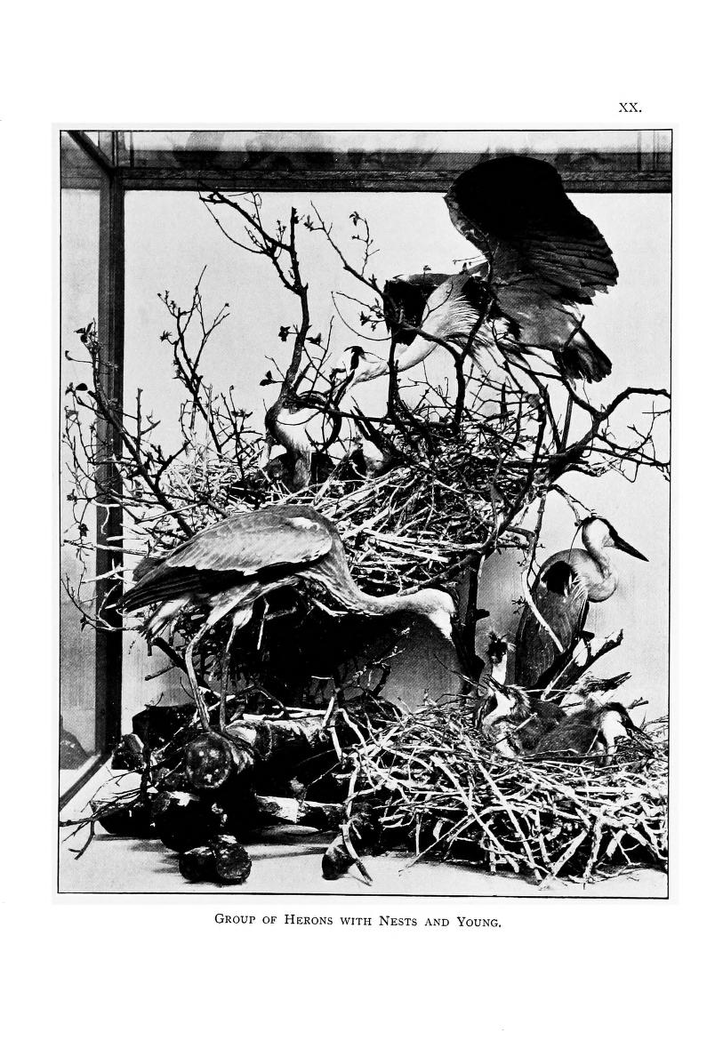
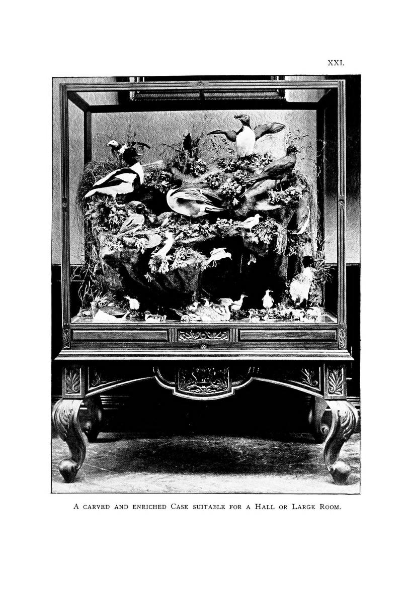
Also part of the taxidermist’s artistry is his or her ability to display the specimens in settings that look like their natural habitat, and to do so in an aesthetically pleasing manner.  Browne treats this subject, and the photographs below are examples of beautifully arranged preserved specimens.

View Full Size Image
Taxidermy of several Herons.

View Full Size Image
Taxidermy of a group of penguins.

Whatever your opinions of taxidermy, it has been crucial to building the collections of many museums we know and love, and to helping our understanding of many different animal species.

July 3, 2013by jjones

Help Support BHL

BHL's existence depends on the financial support of its patrons. Help us keep this free resource alive!

search

About BHL

The Biodiversity Heritage Library (BHL) is the world’s largest open access digital library for biodiversity literature and archives. BHL operates as a worldwide consortium of natural history, botanical, research, and national libraries working together to digitize the natural history literature held in their collections and make it freely available for open access as part of a global “biodiversity community.”

Join Our Mailing List

Sign up to receive the latest news, content highlights, and promotions.

Subscribe Now

Subscribe to Blog via Email

Enter your email address to subscribe to this blog and receive notifications of new posts by email.

Join 319 other subscribers

Subscribe to Blog Via RSS

Subscribe to the blog RSS feed to stay up-to-date on all the latest BHL posts.

Access RSS Feed

Inspiring Discovery through Free Access to Biodiversity Knowledge.

The Biodiversity Heritage Library makes it easier than ever for you to access the information you need to study and explore life on Earth…for free, anytime, anywhere.


64+ Million Pages of
Biodiversity Literature Online.

EXPLORE

Tools and Services
to Transform Research.

EXPLORE

300,000+
Illustrations on Flickr.

EXPLORE

ABOUT | HARMFUL CONTENT | PRIVACY | SITE MAP | TERMS OF USE