

Tasting Platypus Milk: Linking Specimens and Stories
Zoological knowledge typically comes from one of two primary sources: the living and the dead — observations of animals going about their business in their habitats; and the study of preserved specimens. We rarely get the whole picture of an animal’s natural history without both, and each feed into how species are portrayed to those that have never seen them.
Getting echidnas wrong

Taxidermy short-beaked echidna (Tachyglossus aculeatus) from the University Museum of Zoology, Cambridge. Note the backwards-pointing hind feet. Specimen A1. 2/7. Photo Credit: University of Cambridge
Consider the echidna, for example. Examining echidna specimens in museums helps us to understand their evolutionary relationships and enables us to come up with some reasonable suggestions for how they live their lives. The impenetrable coat of thick spines is clearly for defense against predators and guessing at an ant-eating lifestyle doesn’t require too much imagination when you think about what its narrow toothless snout and stout, digging claws might be for. However, we wouldn’t be able to confirm exactly how these things are put to use without seeing them alive.
As someone with one foot in the world of Australian mammal ecology (the living) and another in natural history museums (the dead), one thing that really interests me is the cross-over: how do the dead represent the living? Museums are sites of communication — they provide windows onto the natural world for people to engage with animals they may never see alive. This has made the topic of how animals are depicted — in writings, illustrations and museums — a real focus for me.
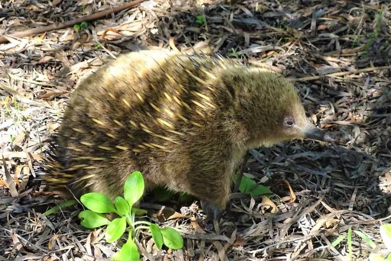
A live (undoctored) echidna, in Tasmania. Photo Credit: Jack Ashby.
While museum specimens are in one sense a primary source, they are also modified by people in order to make them presentable to the public. Taxidermy is intended to make visitors forget the animal is dead, and to achieve that a flat animal skin has to be rebuilt into the shape of a living animal. The trouble is that throughout history, the person doing the taxidermy — particularly for specimens that had travelled to Europe from Australia — had often never seen the living animal. This means that their poses are often incorrect.
In the case of the echidna, it is extremely common for the back feet to point in the wrong direction — in life they should point backwards (enabling echidnas to scratch in between their spines and dig vertically downwards to bury themselves in defense), but taxidermists often didn’t know that, so twisted their feet around. I’ve come across tens of wrong-footed echidnas, and I know the reason why, but I was flummoxed a few years ago when I encountered an echidna with blue eyes at the Manchester Museum. Where did that error come from?
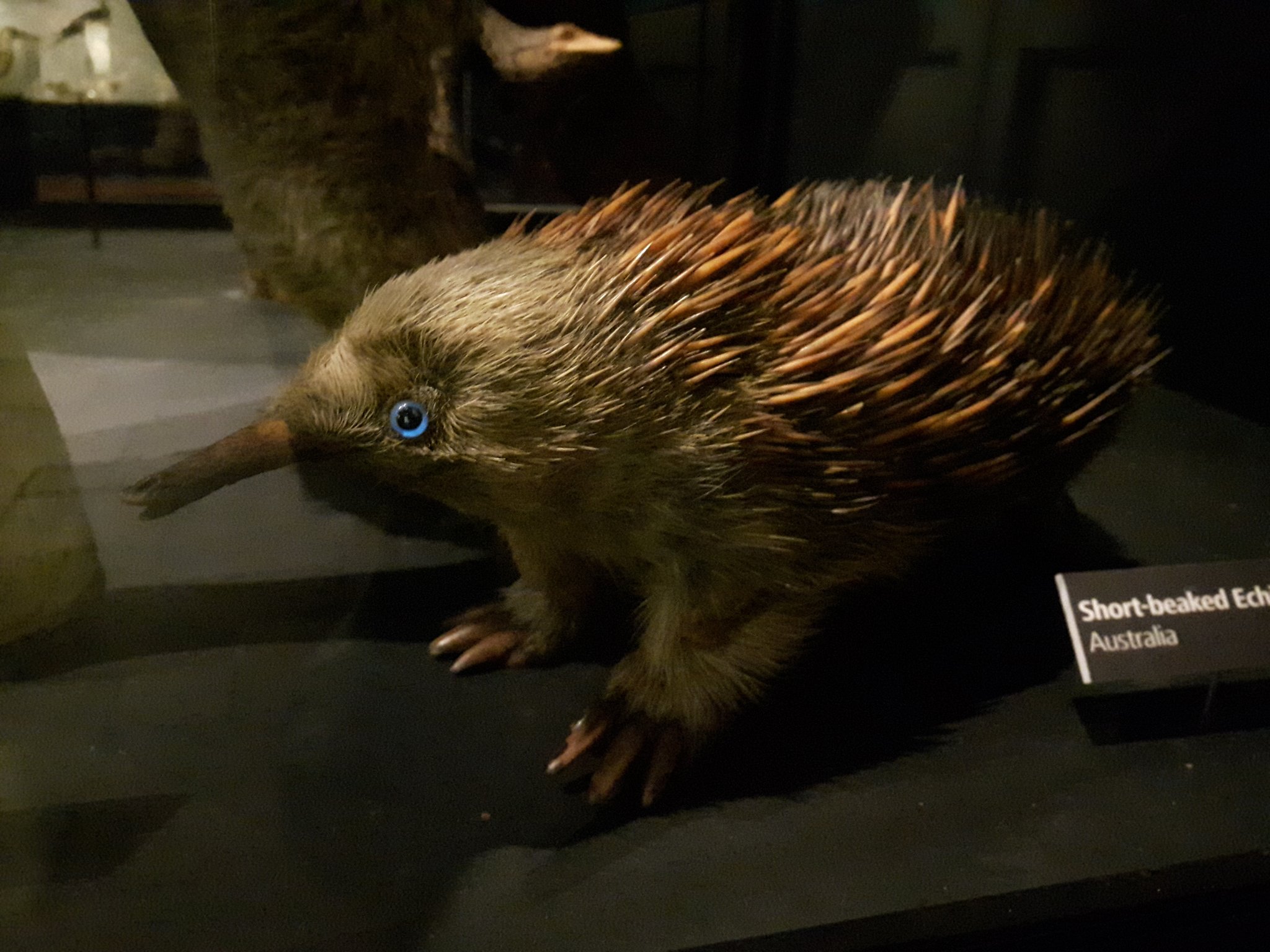
Why does this taxidermy echidna at Manchester Museum have blue eyes? Photo Credit: Jack Ashby.
That mystery echidna popped back into my mind recently when re-reading the original species description on the Biodiversity Heritage Library, and I think I know the answer.
In 1792 George Shaw (who would also publish the first scientific description of the platypus seven year later) included “The Porcupine Ant-Eater” in The Naturalist’s Miscellany. Amongst an otherwise perfectly reasonable description of the species (which he considered to provide a link between actual porcupines in the genus Hystrix and actual ant-eaters in the genus Myrmecophaga), Shaw includes this line:
“The snout is long and tubular, and perfectly resembles in structure that of the Myrmecophaga jubata, or great ant-eater; having only a very small opening or rictus at the tip, from whence is protruded a long lumbriciform [worm-like] tongue, as in the ant-eaters. The nostrils are small, and seated near the extremity of the snout. The eyes are very small, and black, with a pale-blue iris.”
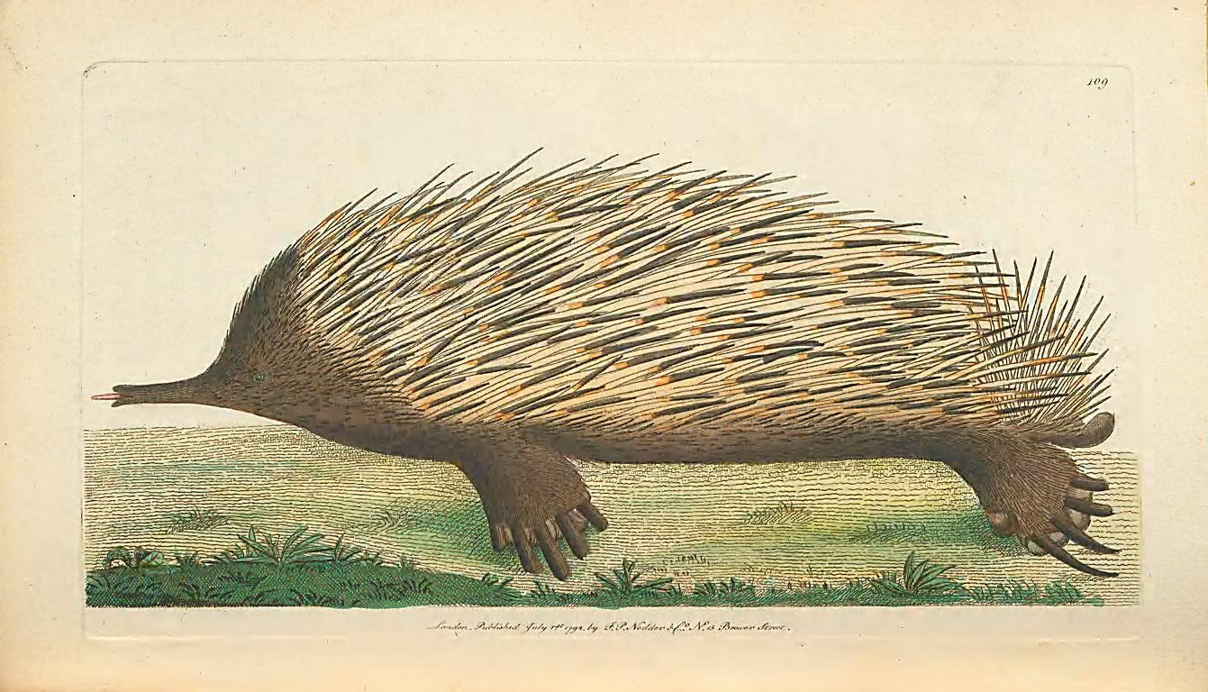
Plate 109. Shaw, George. The naturalist’s miscellany. Volume iii. 1792. Contributed in BHL by Museums Victoria.
Curiously, the illustration that accompanies the text (above) — which is assumed to be based on a painting by Sydney-based collector John White — has correctly coloured black eyes. The question then becomes why Shaw thought echidnas had blue eyes — could it be that the eyes of Shaw’s specimen (sent by Joseph Banks) had been discoloured by the preservative? In any case, it seems likely that the echidna in Manchester was modified to fit Shaw’s original description.
Elsewhere on BHL, we can find examples of illustrations of echidnas that were clearly based on doctored dead specimens, rather than observations of living ones, as their feet incorrectly point forwards and the animals appear flattened. Artists were basing their depictions on “primary sources” in museum collections, but unfortunately those specimens weren’t accurate.
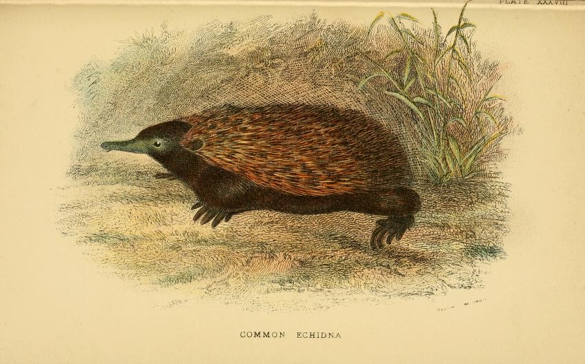
Plate XXXVIII, an echidna with forward-facing feet. Lydekker, Richard, A hand-book to the marsupialia and monotremata. 1896. Contributed in BHL from Smithsonian Libraries.
Tasting platypus milk
I am currently writing a new book on the echidnas’ relative, the platypus. I’ve been utterly reliant on the incredible collection of works on BHL to tell the story of the importance of platypus representations on the history of taxonomy. The introduction of the species to scientific circles in Europe sparked a decades-long debate over where it fitted on the tree of life and what characteristics mammals could possess. One of the many arguments in contention was whether or not they are mammals, another was whether or not they lays eggs. We now know they are, and they do.
A defining feature of mammals is that they feed their young with milk, so those who argued that platypuses are mammals were searching for evidence of milk-producing mammary glands even though platypuses lack nipples. Richard Owen was among those who was attempting to prove their milkiness.
The battle is played out in the documents available on BHL, and to my absolute delight, it led me to this line in one of Owen’s papers on platypus anatomy. Owen had squeezed what he thought was the mammary gland of a preserved platypus and watched the holes where the glands opened onto the skin:
“[T]here escaped from these orifices minute drops of a yellowish oil, which afforded neither perceptible taste nor smell, except such as was derived from the preserving liquor”.
Owen was so dedicated to his argument that he tasted the milk from a platypus that had been dead for many months, or even years.
Without a resource like BHL, putting together the pieces of the platypus puzzle across 220 years, and the changing way that specimens and field-observations have been used to inform representations of the animal over time, would be extremely difficult.
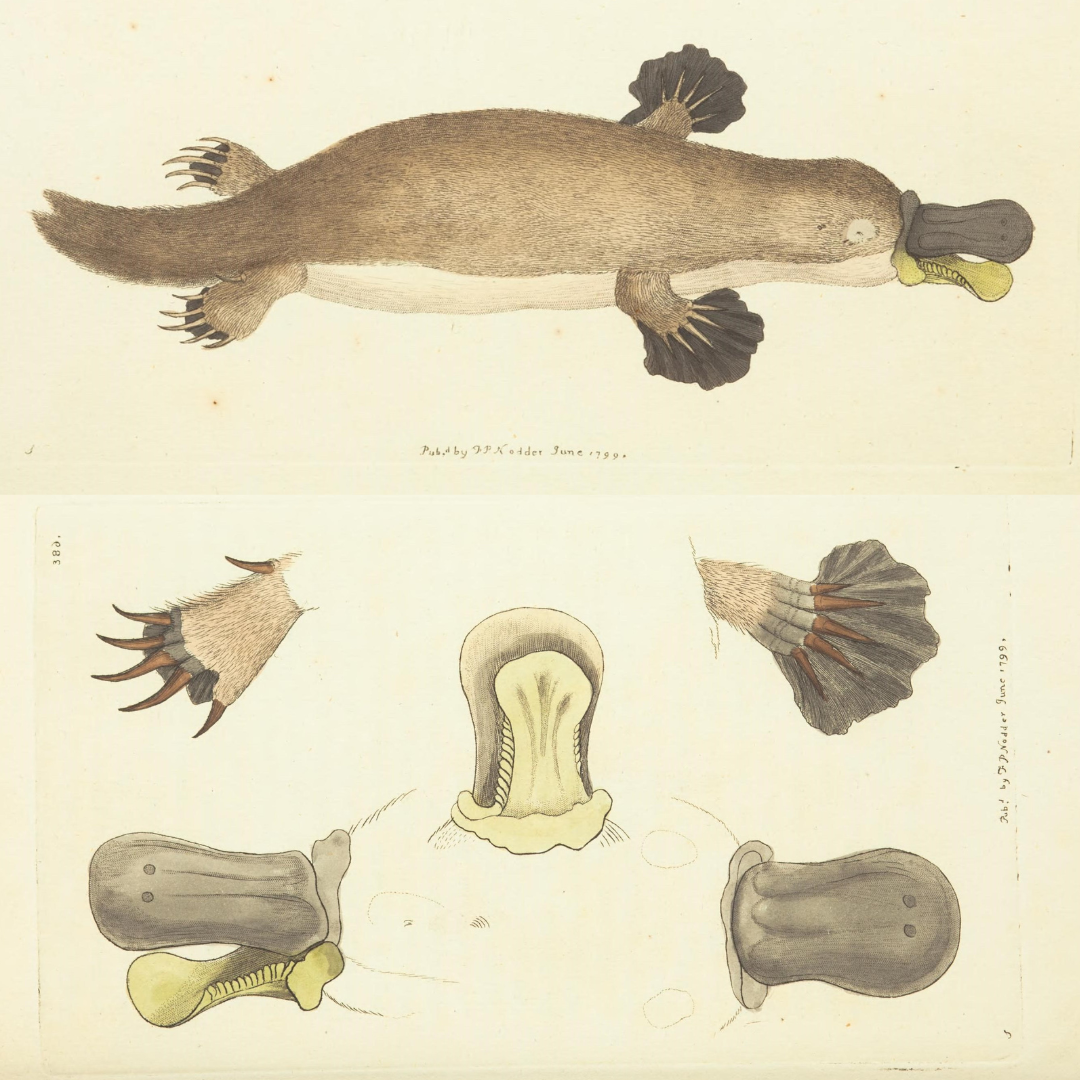
George Shaw, who provided the first scientific description and published illustration of the platypus (Ornithorhynchus anatinus) in 1799 within The Naturalist’s Miscellany, wrote: “… it naturally excites the idea of some deceptive preparation by artificial means …”. Shaw, George. The naturalist’s miscellany. v. 10 (1799). Contributed in BHL from Museums Victoria. DOI: https://doi.org/10.5962/p.304567.
Author and zoologist Jack Ashby is Manager of the University Museum of Zoology, Cambridge. His work centres on engaging people with the natural world through museums. Jack’s book Animal Kingdom: A Natural History in 100 Objects, explores what museum specimens tell us about how evolution works; as well as discussing how natural history museums present a potentially unnatural view of nature. Jack’s zoological passion is the mammals of Australia, where he regularly undertakes ecological fieldwork.
Absolutely fascinating! I am reminded of other illustrations in which the animal or plant depicted is not quite right, particularly with coloration issues that likely relate to preservation methods.